Esophagus Anatomy
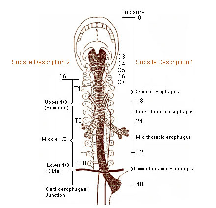
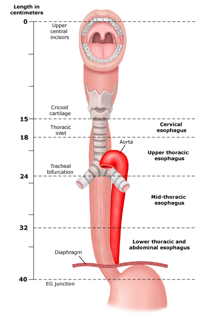
Sub site description 1 cervical. It passes through the muscular diaphragm before entering the stomach.
 Seer Training Anatomy Of The Esophagus
Where the esophagus is compressed by the left main bronchus in the posterior mediastinum.

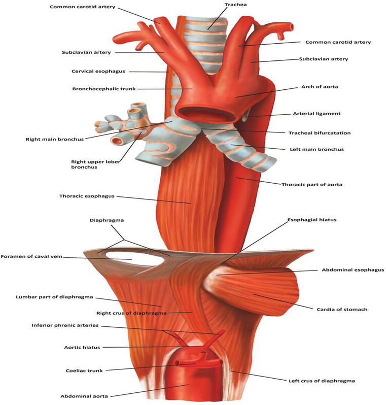
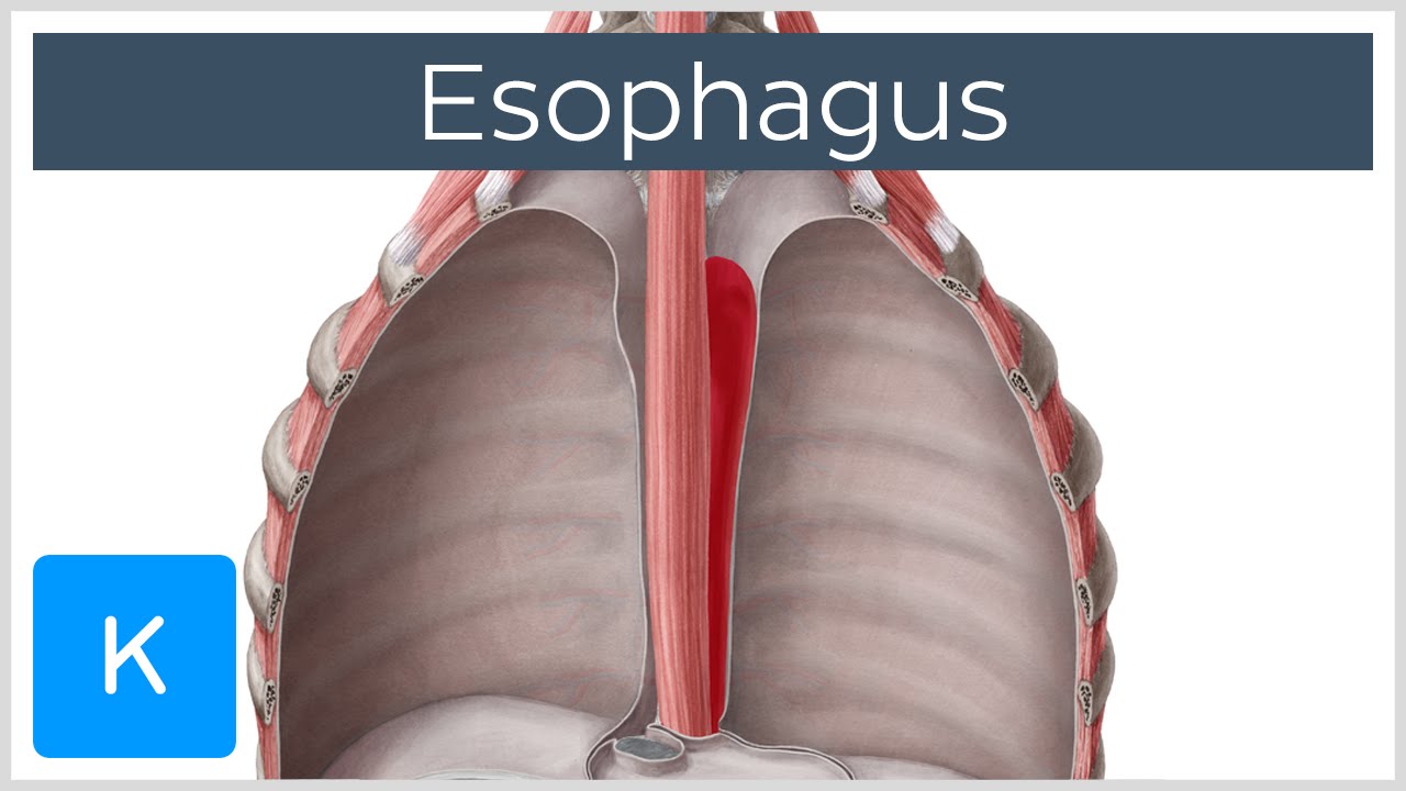
Esophagus anatomy. Gross anatomy relationships of the esophagus. Considered part of lower thoracic esophagus. Where it is crossed on the front by the aortic arch in the superior mediastinum.
In this article we shall examine the anatomy of the oesophagus its structure. Cervical begins at the lower end of pharynx. Anatomically it lies behind the trachea and heart and in front of the spinal column.
This article will highlight the main anatomical features of the esophagus including its constrictions and sphincters its histological layers and the main pathological changes that may ail this particular organ. Venous blood from the esophagus drains into a submucosal plexus. The esophagus is about 8 inches long and is lined by moist pink tissue called mucosa.
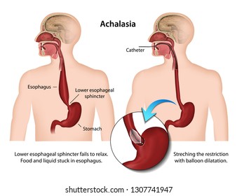
Esophagus also spelled oesophagus relatively straight muscular tube through which food passes from the pharynx to the stomach. At the start of the esophagus where the laryngopharynx joins the esophagus. It originates at the inferior border of the cricoid cartilage c6 extending to the cardiac orifice of the stomach t11.
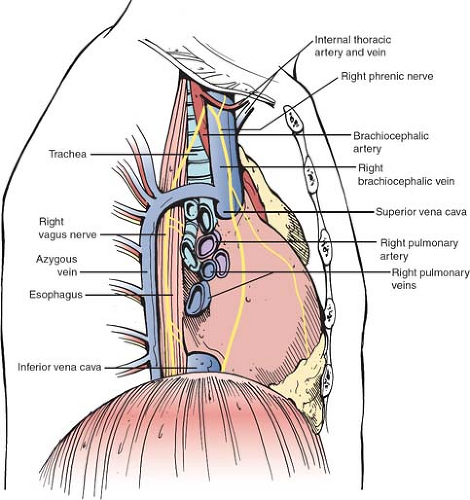
The esophagus is a long thin and muscular tube that connects the pharynx throat to the stomach. The esophagus has 2 types of lymphatic vessels. The trachea lies anterior to the esophagus.
The esophagus can contract or expand to allow for the passage of food. The oesophagus is a fibromuscular tube approximately 25cm in length that transports food from the pharynx to the stomach. It forms an important piece of the gastrointestinal tract and functions as the conduit for food and liquids that have been swallowed into the pharynx to reach the stomach.
The esophagus runs behind the windpipe trachea and heart and in front of the spine. The esophagus is a muscular tube that conveys food and fluids from the pharynx to the stomach. The esophagus is a long fibromuscular tube that runs in the thoracic cavity and connects the pharynx with the stomach.
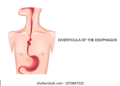
The esophagus is a muscular tube connecting the throat pharynx with the stomach. The esophagus is a hollow muscular tube that transports saliva liquids and foods from the mouth to the stomach. Just before entering the stomach the esophagus passes through the diaphragm.
From thoracic inlet to level of tracheal bifurcation.
 Esophagus Abdominal Key
 Anatomy Of Esophagus Intechopen
 The Anatomy Of The Esophagus Basicmedical Key
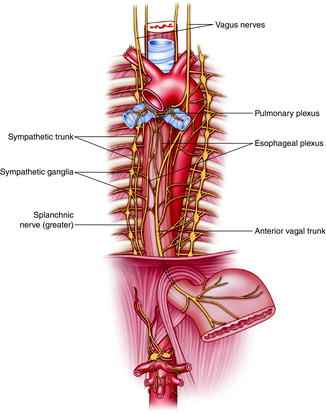
![]() Esophagus Anatomy Sphincters Arteries Veins Nerves Kenhub
Esophagus Anatomy Sphincters Arteries Veins Nerves Kenhub
 Anatomical Relations Esophagus Anatomy
 Surgical Anatomy Of The Esophagus And Esophagogastric
 Esophagus Images Stock Photos Vectors Shutterstock
 Esophagus Pain Neck
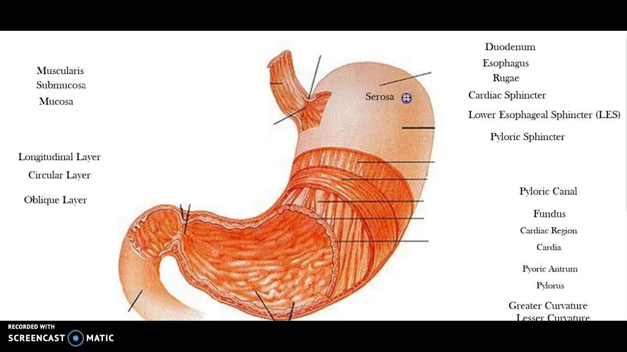 Esophagus And Stomach Anatomy Youtube
 Esophagus Images Stock Photos Vectors Shutterstock
 Musculature Of Esophagus Anatomy Thyroid Cartilage Cricoid
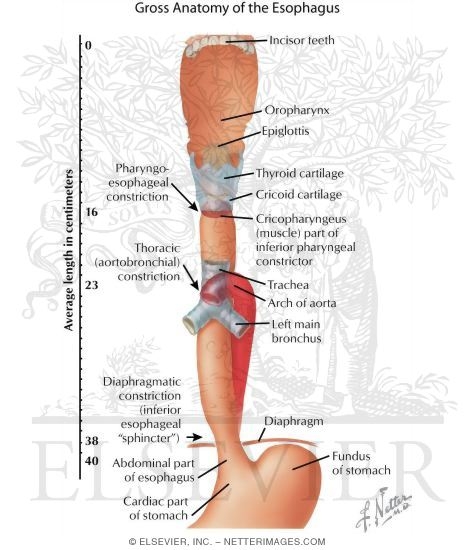 Gross Anatomy Of The Esophagus
![]() Esophagus Anatomy Sphincters Arteries Veins Nerves Kenhub
Esophagus Anatomy Sphincters Arteries Veins Nerves Kenhub

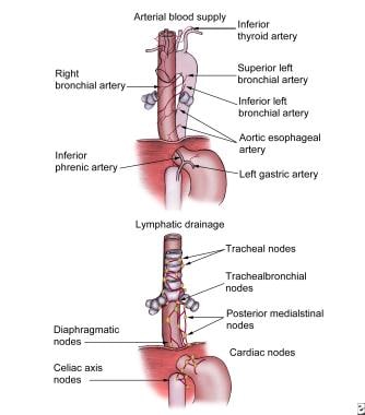 How Is Blood Supplied To The Esophagus In The Anatomy Of
 Esophagus Anatomy
 Adult Cardiothoracic Surgery Esophageal Cancer

 Esophagus Definition Function And Structure Human Anatomy Kenhub
 Esophagus Absite Slayer Accesssurgery Mcgraw Hill Medical
![]() Esophagus Stock Illustrations 4 303 Esophagus Stock
Esophagus Stock Illustrations 4 303 Esophagus Stock
 Development Anatomy And Physiology Of The Esophagus
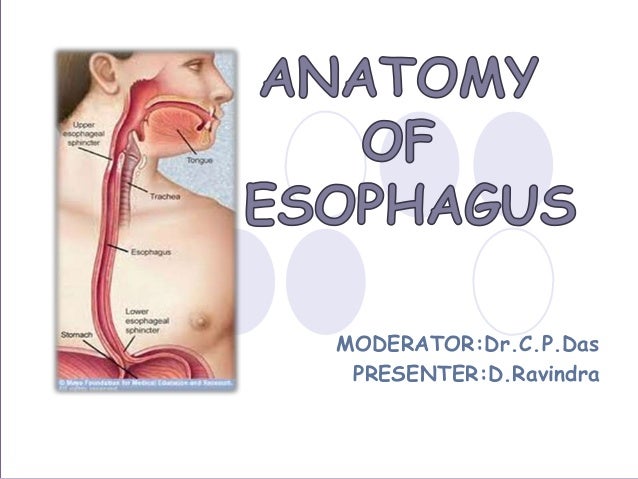 Anatomy Of Esophagus By Dr Ravindra Daggupati
Esophagus Blood Supply Cervical Esophagus Anatomy Gallery
Belum ada Komentar untuk "Esophagus Anatomy"
Posting Komentar