Anatomy Of The Spinal Cord
A component of the central nervous system it sends and receives information between the brain and the rest of the body. It is covered by the three membranes of the cns ie the dura mater arachnoid and the innermost pia mater.
 The Spinal Cord Injury Anatomy Simplified
The spinal cord is a cylindrical structure greyish white in colour.

Anatomy of the spinal cord. Like the brain the spinal cord is covered by three connective tissue envelopes called the meninges. Spinal nerves are grouped as cervical c1 c8 thoracic t1 t12 lumbar l1 l5. Spinal cord anatomy in the neck internal anatomy of the spinal cord.
Cervical spinal cord injury. Lumbar enlargement corresponds to the lumbosacral plexus nerves which innervate the lower limb. The spinal cord arises cranially as a continuation of the medulla oblongata part of the brainstem.
The space between the outer and middle envelopes is filled with cerebrospinal fluid a clear. The spinal cord is part of the central nervous system cns. Cross sectional anatomy of spinal cord.
It has a relatively simple anatomical course. Signs and symptoms of spinal cord compression. Spinal nerve cells called neurons carry messages to and from the spinal cord via spinal nerves.
Spinal cord anatomy the spinal cord is a bundle of nerve fibers that extend from the brain stem down the spinal column to the lower back. Gray matter has a relatively dull color because it contains little myelin. Spinal cord anatomy basically spinal cord is a long and narrow bundle of nervous tissues and support cells which extends from the base of our brain to the upper lumbar region.
The spinal cord is part of the central nervous system cns which extends caudally and is protected by the bony structures of the vertebral column. It passes through the spinal canal or spinal cavity of the vertebral column ie the backbone or spine. There are two regions where the spinal cord enlarges.
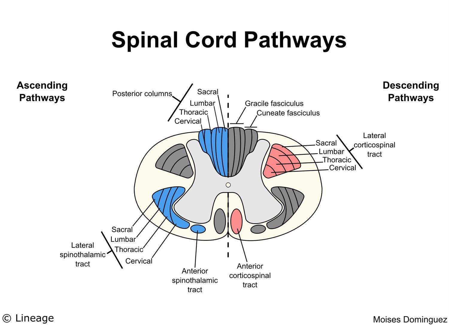
Spinal cord neural pathways are found within the spinal cord white matter. When viewed as a cross section from above. It is composed of nerve fibres that mediate reflex actions and that transmit impulses to and from the brain.
The main job of the spinal cord is to be the communication system between the brain and the body by carrying messages that allow people to move and feel sensation. Protective layers of the spinal cord. It contains the somas dendrites and proximal parts of the axons of neurons.
Cervical enlargement corresponds roughly to the brachial plexus nerves. Anatomy and physiology of the spinal cord. The spinal meninges help prevent the spinal cord.
It then travels inferiorly within the vertebral canal surrounded by the spinal meninges containing cerebrospinal fluid. The spinal cord like the brain consists of two kinds of nervous tissue called gray and white matter.
 Spinal Cord Function Anatomy Physiology Medisavvy
 Spinal Cord Anatomy Parts And Spinal Cord Functions
 13 2 Gross Anatomy Of The Adult Spinal Cord Diagram Quizlet
 The Spinal Cord Boundless Anatomy And Physiology
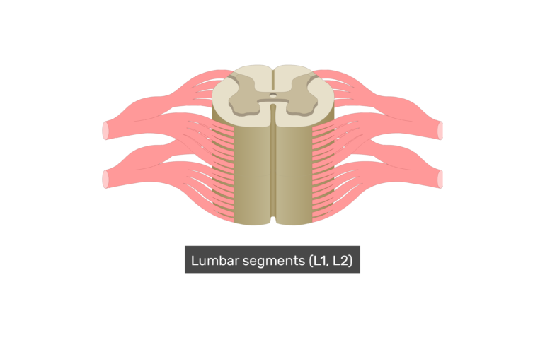 Spinal Cord Segments Cross Sectional Anatomy
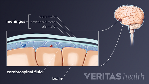 Spinal Cord Anatomy In The Neck
Ch 12 Gross Anatomy Of The Spinal Cord
 The Spinal Cord Human Anatomy And Physiology Lab Bsb 141
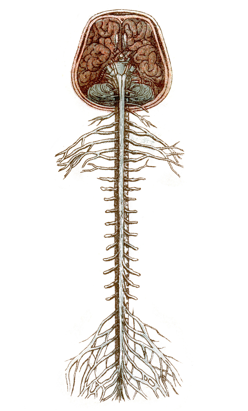 The Spinal Cord Queensland Brain Institute University Of
 Spinal Anatomy Center Cervical Thoracic And Lumbar Spine
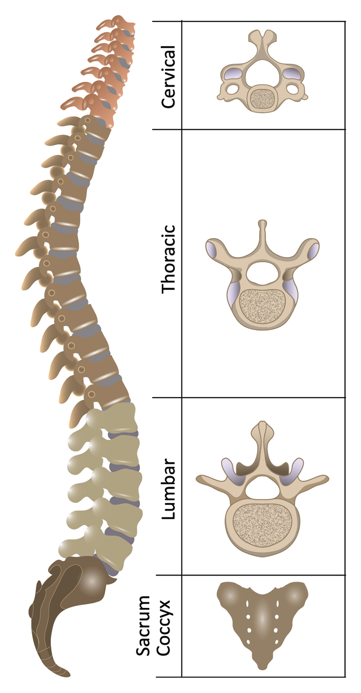 Anatomy Of The Spine Spinal Cord Injury Information Pages
:watermark(/images/watermark_5000_10percent.png,0,0,0):watermark(/images/logo_url.png,-10,-10,0):format(jpeg)/images/atlas_overview_image/456/lu5kV9M3yLK6FA6RJoGpew_spinal-membranes-and-nerve-roots_english.jpg) Spinal Cord Anatomy Structure Tracts And Function Kenhub
Spinal Cord Anatomy Structure Tracts And Function Kenhub
 Spinal Cord Summary Neuroanatomy Geeky Medics
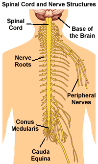 Understanding Spinal Anatomy Spinal Cord And Nerve Roots
 Spinal Cord Anatomy In The Neck
 Anatomy Of The Spinal Cord Download Scientific Diagram
 Spinal Cord Injury Anatomy And Physiology Of The Spinal Cord
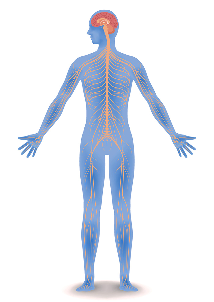 Central Nervous System Brain And Spinal Cord Queensland
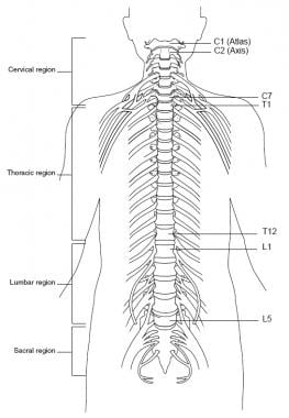 Topographic And Functional Anatomy Of The Spinal Cord Gross
 Spinal Cord Lesions Neurology Medbullets Step 2 3
 Game Statistics Close Up Anatomy Spinal Cord Purposegames
Anatomy Of Spinal Blood Supply
Belum ada Komentar untuk "Anatomy Of The Spinal Cord"
Posting Komentar