Pancreas Anatomy Ultrasound
In most cases the transversal and sagittal directions. The pancreas is an oblong shaped organ positioned at the level of the transpyloric plane.
 Startradiology
Preparation fast the patient to reduce interference from overlying bowel gas which may otherw.
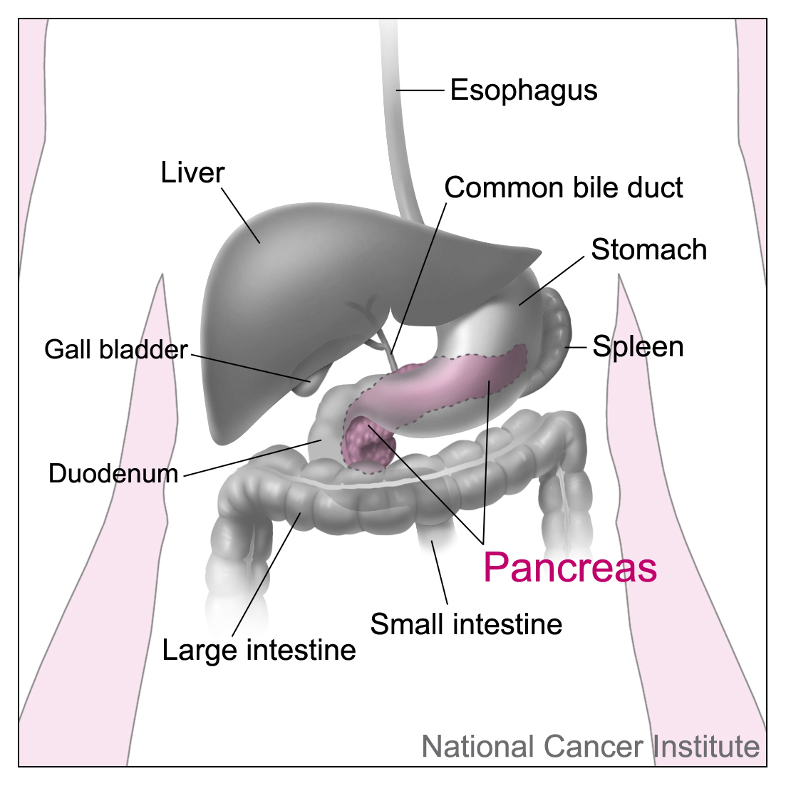
Pancreas anatomy ultrasound. It is involved in the production of hormones insulin glucagon and somatostatin and also involved in digestion by its production and secretion of pancreatic juice. 202 pylorus pancreas liver. The head of the pancreas is drained by the two anterior and posterior inferior pancreaticoduodenal veins which empty into the superior mesenteric vein.
December 17 2002 by lars thorelius. The pancreas is a retroperitoneal organ that has both endocrine and exocrine functions. Ultrasound of the pancreas.
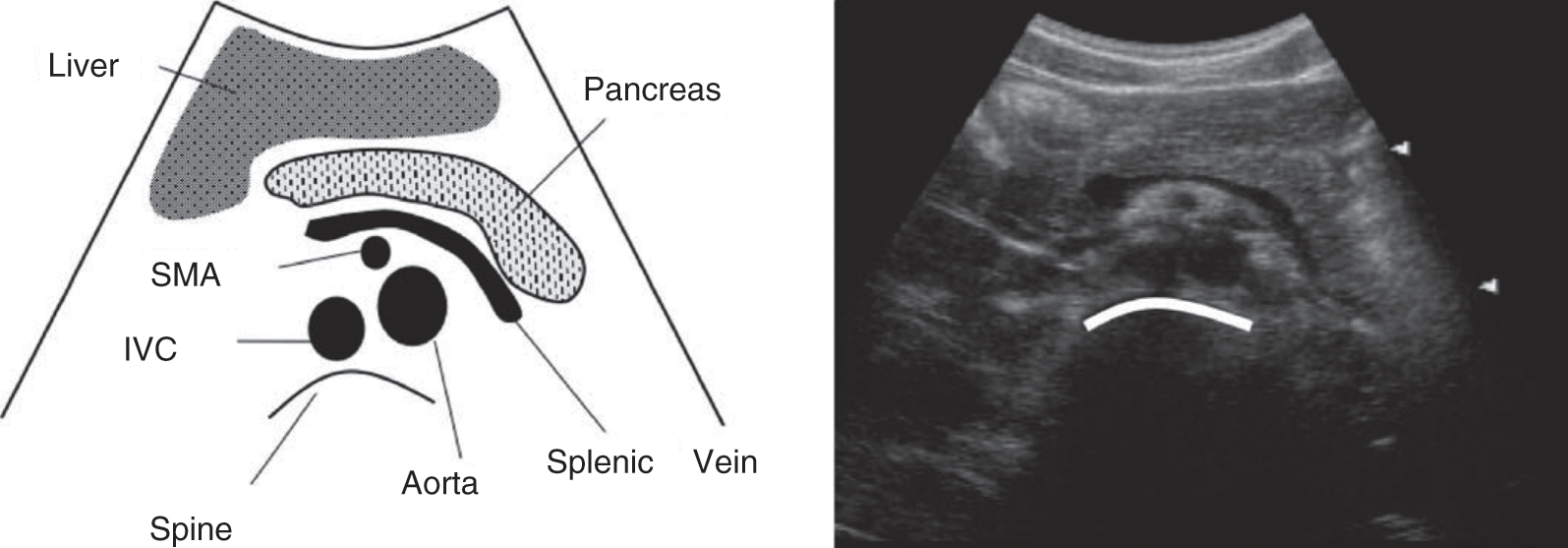
Use the splenic vein to help identify the pancreas superficial to this. A common sequence of a full abdominal ultrasound examination is aorta pancreas livergallbladder kidneys bladder region intestines. Ultrasound evaluation of the normal pancreas cme vital activity provides an overview of normal pancreas anatomy structural and vasculature frames as well as the lab values indicating pancreatic disease.
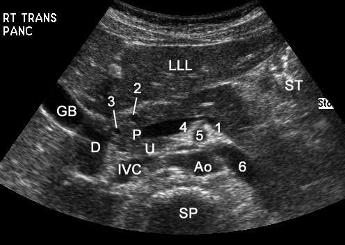
The anterior and posterior superior pancreaticoduodenal veins drain directly into the portal vein. Pancreatic ultrasound can be used to assess for pancreatic malignancy pancreatitis and its complications as well as for other pancreatic pathology. Tail of pancreas start with the probe transverse then angle the heel of the probe cephalad and left as the tail can be sitting up under the spleen.
As a general rule each organ and abnormality is imaged in two directions. The pylorus is characterized by a marked thickening of the muscular coat anterior to the head of the pancreas. With the exception of the tail of the pancreas it is a retroperitoneal organ located deep within the upper abdomen in the epigastrium and left hypochodrium regions.
However the complex anatomy of the organ and surrounding tissues make evaluation a demanding task and the ultrasound echo of even the normal pancreas varies widely from patient to patient. A longitudinal scan through the upper midabdomen demonstrates the characteristic triad of stomach liver and pancreas. The location of the pancreas in the abdomen makes it well suited for ultrasound examination.
Thus the spleen can be used as a window and a left intercostal coronal approach can also be utilised.
 Transabdominal Ultrasonography Of The Pancreas Basic And
 Presentation1 Pptx Ultrasound Study Of The Spleen And Pancreas
 Endoscopic Ultrasound And Pancreatic Disorders Tenet
 Chapter 8 Ultrasound Of The Pancreas Surgical And
 Right Upper Quadrant Ultrasonography Chapter 5 Atlas Of
 Ultrasound Of The Pancreas
 Ultrasound Of The Pancreas What Normal Looks Like
 Roadmap Developed For Role Of Focused Ultrasound In Treating
 Abdominal Ultrasound Chapter 24 Clinical Emergency Radiology
 Pancreas Function Location Diseases Live Science
 Ultrasound Nick S Radiology Wiki
 Anatomy Of The Pancreas And Surrounding Organs Diagram
![]() The Pancreas Anatomy Duct System Vasculature
The Pancreas Anatomy Duct System Vasculature
 What Is A Pancreatic Cyst Everyday Health
 Abdominal Ultrasound Registry Review
 Pancreas An Overview Sciencedirect Topics
 Longitudinal View Of Pancreas
 Normal Pancreas Ultrasound How To
 Pancreatic Sonographic Anatomy
 Computed Tomography Ct Scan Of The Pancreas Johns
 Relation Of Ultrasound Findings And Abdominal Symptoms
 Pancreatic Sonographic Anatomy
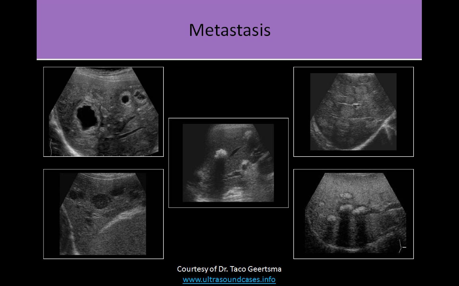
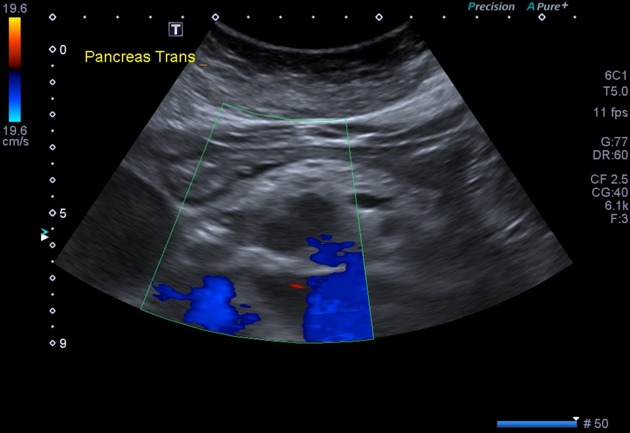
Ultrasound Of Pancrease In Radiology
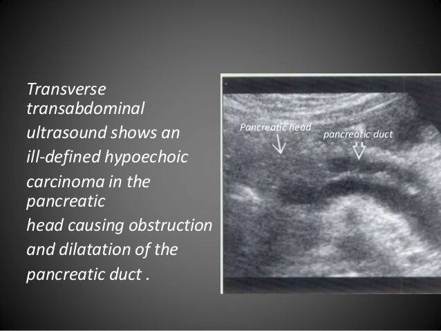
 Pancreatic Adenocarcinoma Radiology Case Radiopaedia Org
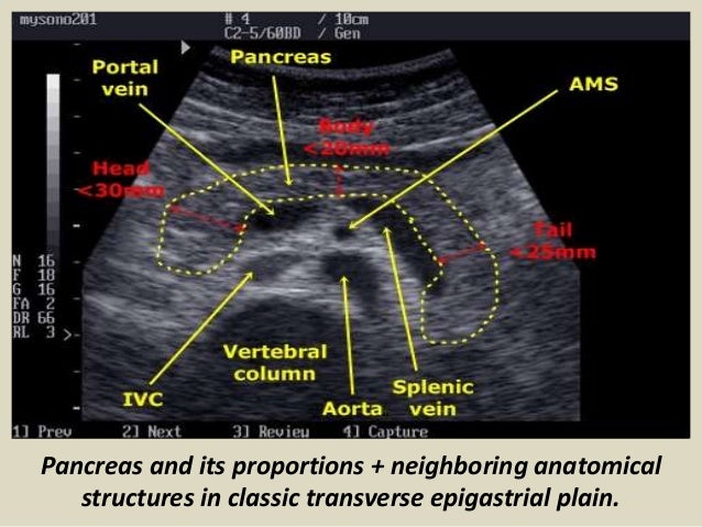
 Liver Measurement Ultrasound Pancreas And Its Proportions


Belum ada Komentar untuk "Pancreas Anatomy Ultrasound"
Posting Komentar