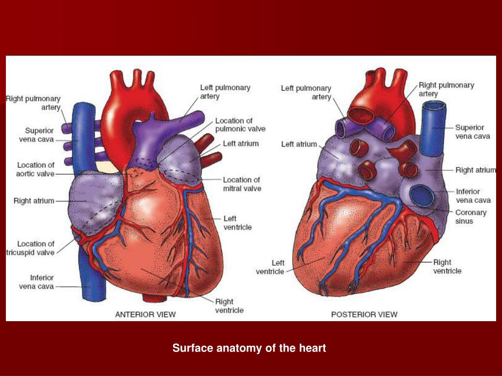
Surface Of Heart Anatomy
Surface anatomy of heart and lung dr. Fig 10 borders of the heart.
 Surface Anatomy Of Heart And Lungs
In birds this is termed topography.

Surface of heart anatomy. Surface anatomy deals with anatomical features that can be studied by sight without dissection. Heart valves separate atria from ventricles and ventricles from great vessels. The heart is a hollow structure.
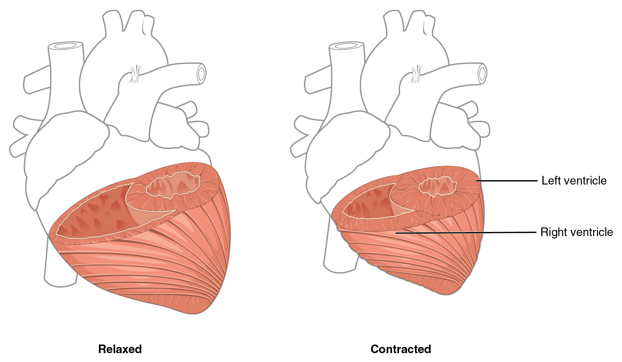
Chambers of the heart. Video created by rob swatski associate professor of biology harrisburg area community college york campus york pa. Made up of the surface of the left ventricle right pulmonary surface.
Examining the surfaces of the heart. The heart is composed of 4 chambers right atrium and right ventricle and left atrium and left ventricle. The base of the heart is located at the level of the third costal cartilage as seen in figure 1.
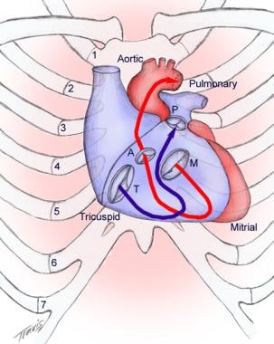
Blood flow through the heart. Mohamed el fiky professor of anatomy and embryology. The blood flow through the heart is quite logical.
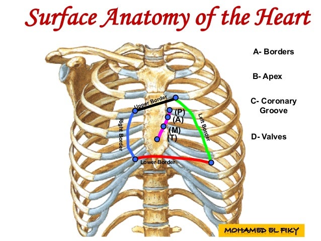
Separating the surfaces of the heart are its borders. Surfaces and borders of the heart orientation and surfaces. Surface anatomy also called superficial anatomy and visual anatomy is the study of the external features of the body of an animal.
Made up by the surface of the right atrium the heart has four ausculatory areas. A web of nerve tissue also runs through the heart conducting the complex signals that govern contraction and relaxation. On the surface the atria are divided from the ventricles by the atrioventricular groove also named coronary sulcus and ventricles from every other by interventricular grooves.
As such it is a branch of gross anatomy. Sulci of the heart. The heart has been described by many texts as a pyramid which has fallen.
Blood supply of the heart. Surface anatomy of heart and lungs 1. Location of the heart.
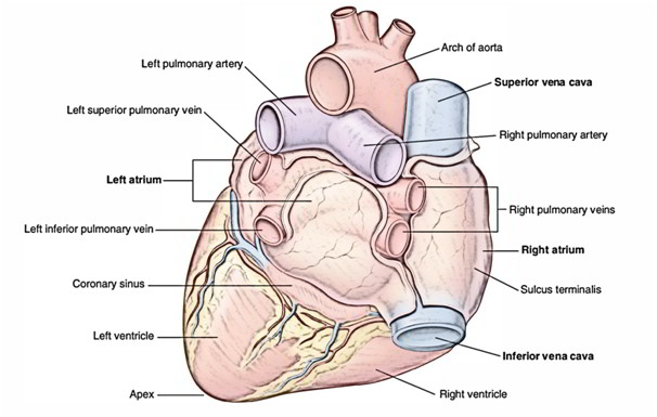
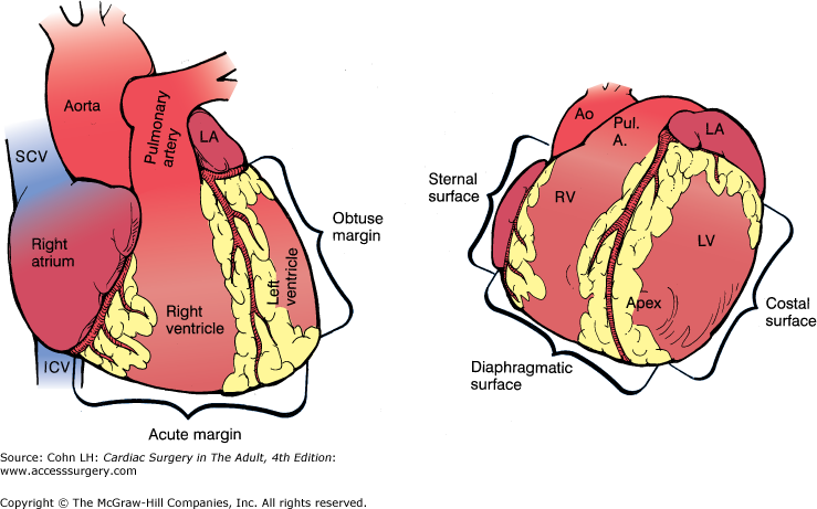
The heart has five surfaces. Surrounding the heart is a sac called the pericardium. The inferior portion formed by the left ventricle and part of the right ventricle left pulmonary surface.
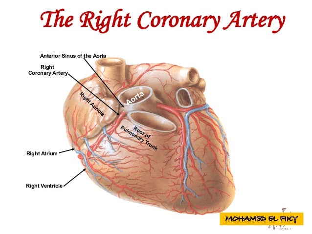
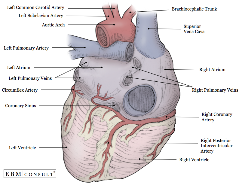
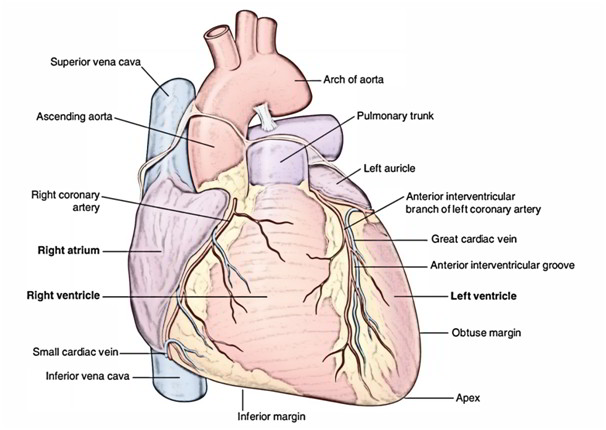
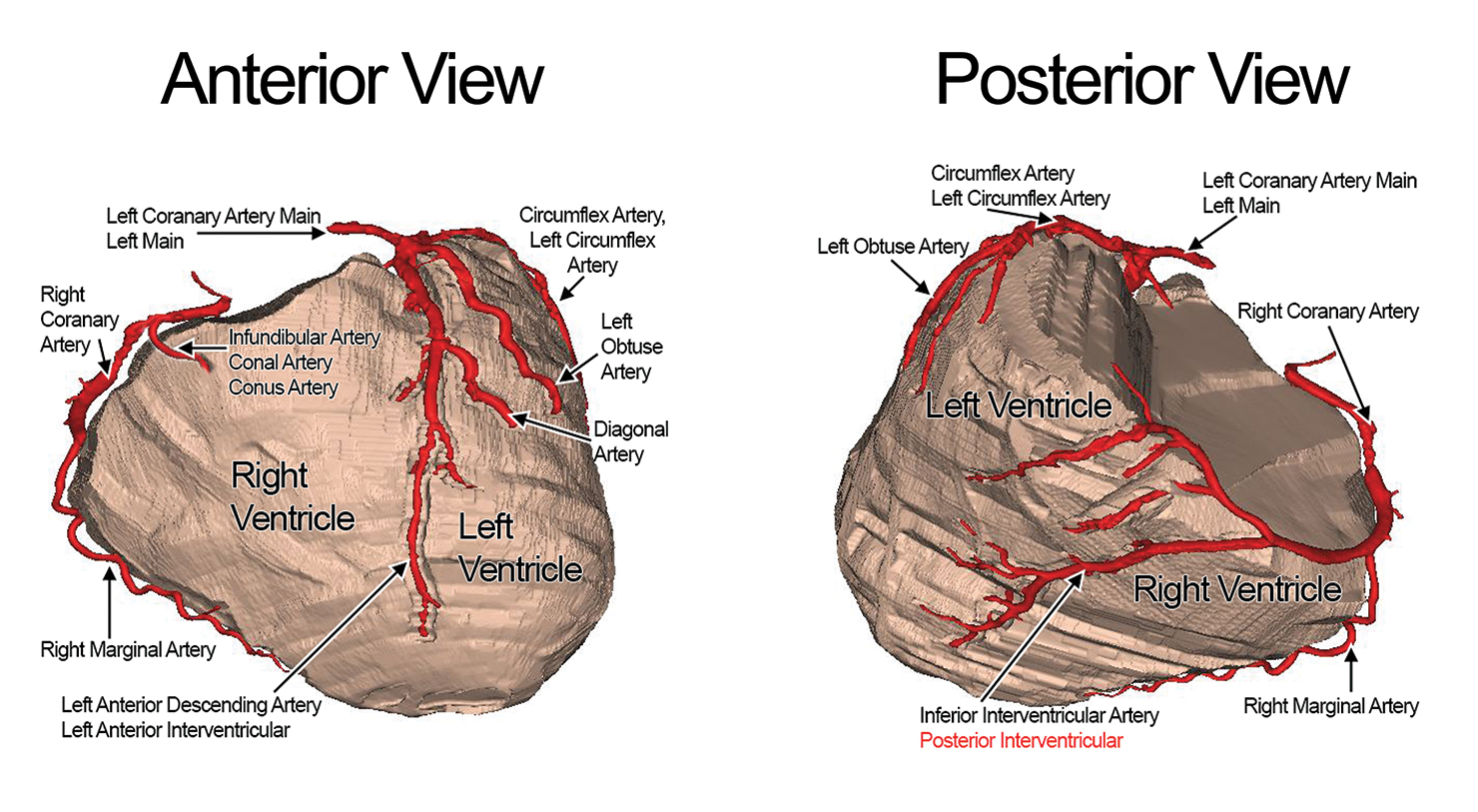
As the heart contracts it pumps blood around the body. The great veins the superior and inferior venae cavae and the great arteries the aorta and pulmonary trunk are attached to the superior surface of the heart called the base. Right coronary artery right atrium right ventricle anterior sinus of.
It sits in the chest slightly to the left of center. A review of the external anatomy of the heart and the great vessels. The heart is a muscular organ roughly the size of a closed fist.
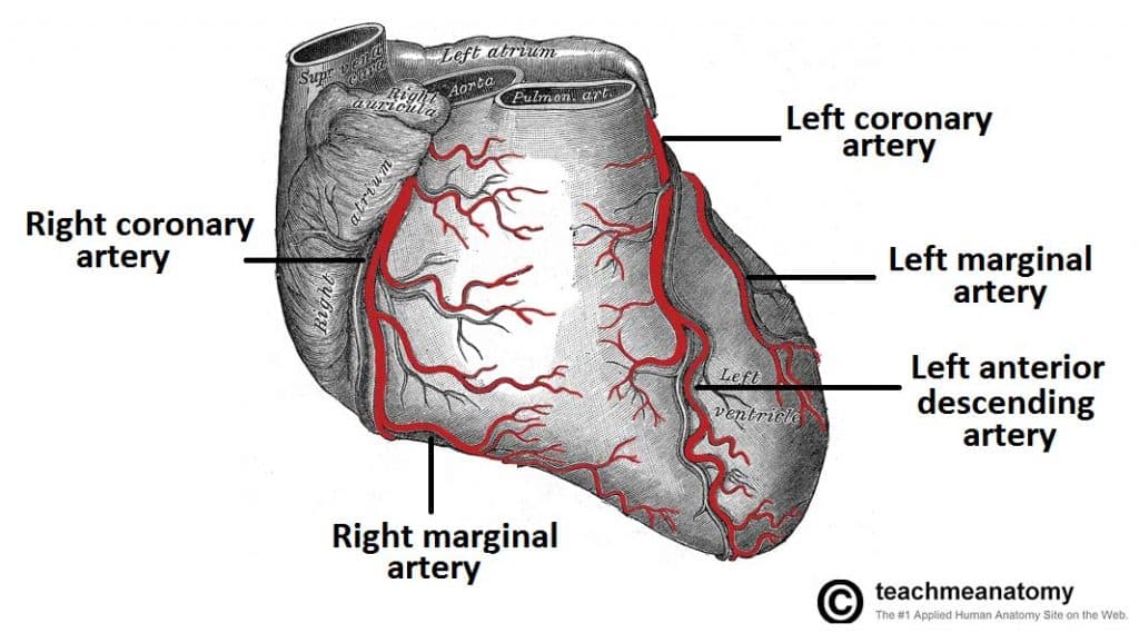
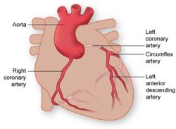
Base posterior diaphragmatic inferior. The coronary arteries run along the surface of the heart and provide oxygen rich blood to the heart muscle. Right atrium right ventricle left coronary artery right.
 Easy Notes On Heart Learn In Just 4 Minutes Earth S Lab
 2 Coronary Vessels Supplying And Draining Blood To The
 Anatomy 1 C1 L8 Surfaces Of The Heart
 The Heart
 Vasculature Of The Heart Teachmeanatomy
 Anatomy Heart External
 Heart Structure Anatomy Physiology Wikivet English
 Chapter 2 Surgical Anatomy Of The Heart Cardiac Surgery
 Coronary Arteries Texas Heart Institute
 6 The Heart
Circulatory System Anatomy Physiology 1224 With Rooney
How The Heart Works Saint Luke S Health System
 Easy Notes On Heart Learn In Just 4 Minutes Earth S Lab
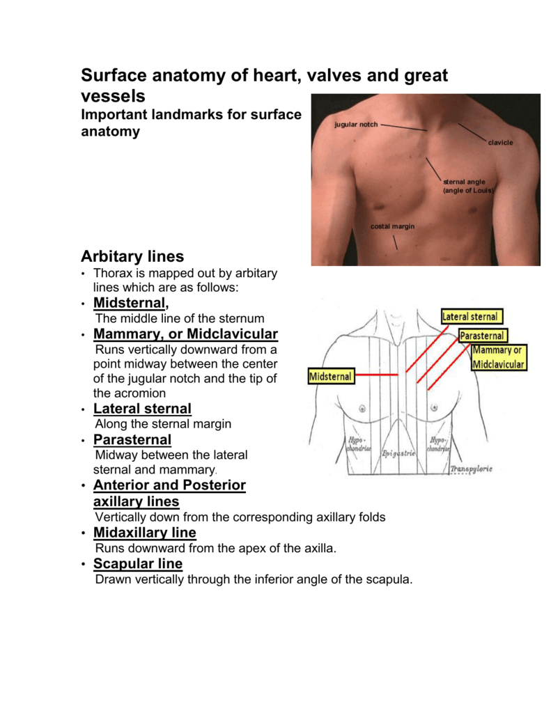 Surface Anatomy Of Heart Valves And Great Vessels
 Surface Anatomy Of Heart And Lungs
 19 1 Heart Anatomy Anatomy And Physiology
 Atrium Heart Wikipedia
 Surface Anatomy Heart Failure Guws Medical
 Surface Anatomy Wikipedia
 Heart Anatomy Anatomy And Physiology Ii
 Pediagenosis
 Heart External Features Anatomy Qa
 Tricuspid Valve Anatomy Overview Gross Anatomy
 Coronary System Tutorial What Is The Coronary System
 Ppt Assessment Of The Cardiovascular System Powerpoint
 Surface Anatomy Of Heart Madeline M Lee
 Heart Anatomy Anatomy And Physiology Ii

Belum ada Komentar untuk "Surface Of Heart Anatomy"
Posting Komentar