Hamstring Muscles Anatomy
Grade 1 strains include milder strains that can be treated at home. These muscles start at the bottom of your pelvis extending down the back of your thigh and along either side of your knee to your lower leg bones.
 Your Physical Therapy Home Exercises To Strength Hamstring
Muscles will be innervated by the tibial branch of the sciatic nerve.

Hamstring muscles anatomy. The common criteria of any hamstring muscles are. Grade 2 strains are more severe and include more loss of range of motion. The hamstring muscles the hamstring muscles cross two joints the hip and the knee and can act as extensors of the thigh and flexors of the leg.
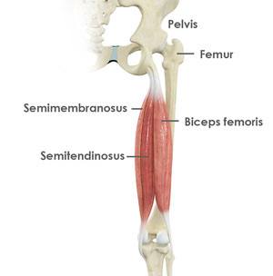
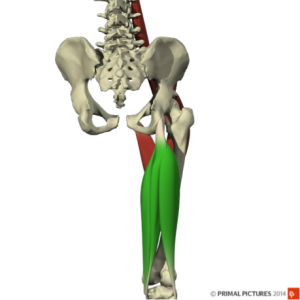
The muscles in the posterior compartment of the thigh are collectively known as the hamstrings. The hamstrings are comprised of three separate muscles. Anatomy of the hamstring muscles.
Working the muscle also creates strength and toughness in the tendons that attach the muscle to the bone making them less likely to strain and tear. As group these muscles act to extend at the hip and flex at the knee. Muscle will participate in flexion of the knee.
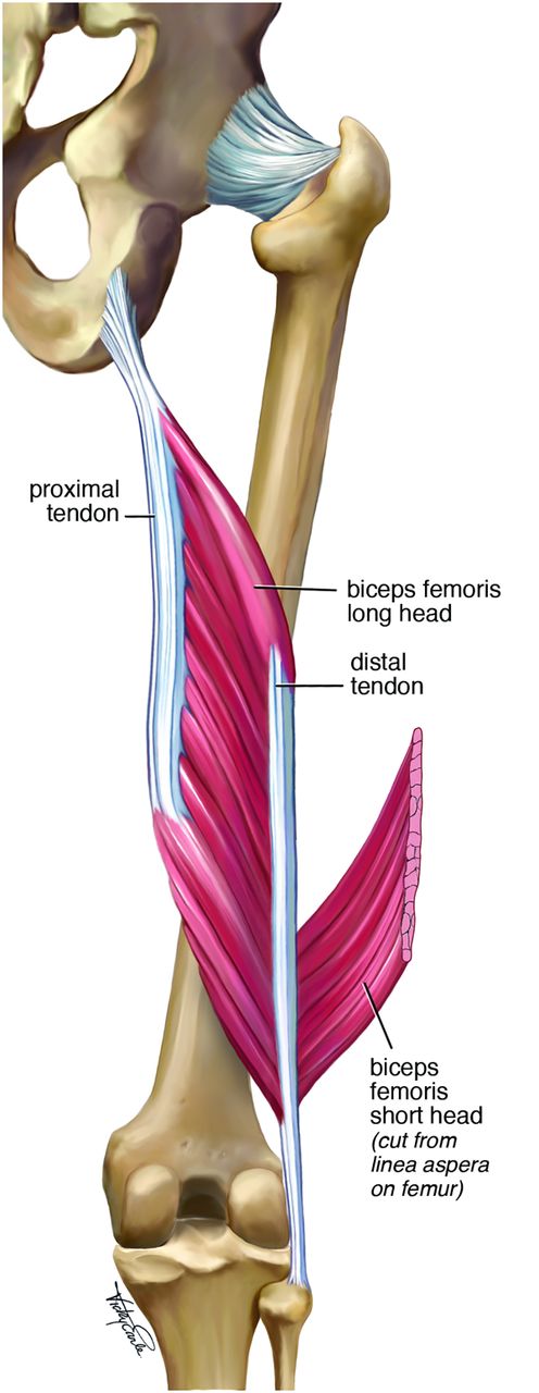
Anatomy of the hamstring muscles. Like the biceps in your upper arm the largest of the hamstring muscles biceps femoris has two heads long and short. On the other hand hamstring strengthening poses are typically practiced less often so were missing out on their ability to build endurance in the actual muscle fibers.
Muscles should be inserted over the knee joint in the tibia or in the fibula. There are three grades of hamstring strain. The hamstring tendons flank the space behind the knee.
The biceps femoris semitendinosus and semimembranosus. The hamstrings refer to 3 long posterior leg muscles the biceps femoris semitendinosus and semimembranosus. They consist of the biceps femoris semitendinosus and semimembranosus which form prominent tendons medially and laterally at the back of the knee.
The biceps femoris is the only two headed hamstring muscle. These muscles originate just underneath the gluteus maximus on the pelvic bone and attach on the tibia. The largest muscle of the hamstrings is the biceps femoris.
It starts at the bottom of the pelvic bone. Because these muscles have a common site of origin on the ischial tuberosity and have similar positions in the posterior thigh. The long head is the one toward the outside of the thigh.
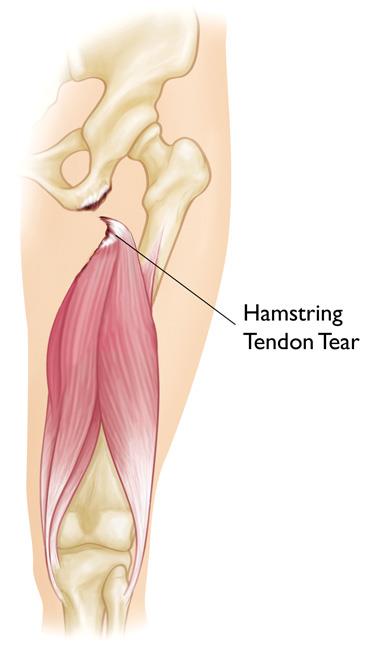
The hamstring muscles have their origin where their tendons attach to bone at the ischial tuberosity of the hip often called the sitting bones and the linea aspera of the femur. Severe grade 3 strains may include avulsion where some part of the muscle actually detaches from its connection to bone. The most medial muscle the semimembranosus.
Muscles should originate from ischial tuberosity.
 Hamstring Tendonitis Or Hamstring Syndrome Zion Physical
 Strength Is Never A Weakness Hamstring Heaven
 Pin By Lindsay Gallagher On A Life Time Of Sports Injuries
 The Mighty Hamstring Muscles Anatomy Injury Training
 Muscles Advanced Anatomy 2nd Ed
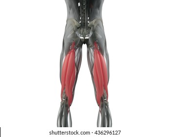 Hamstring Muscle Anatomy Images Stock Photos Vectors
 Proximal Lesion A Schematic Representation Of The
 Muscle Anatomy Of The Semitendinosus
 Hamstring Muscles 3d Anatomy Tutorial
 Anatomy 101 Understanding Your Hamstrings Hamstring
 Semitendinosus Muscle Wikipedia
 Anatomy Tutorial The Hamstrings
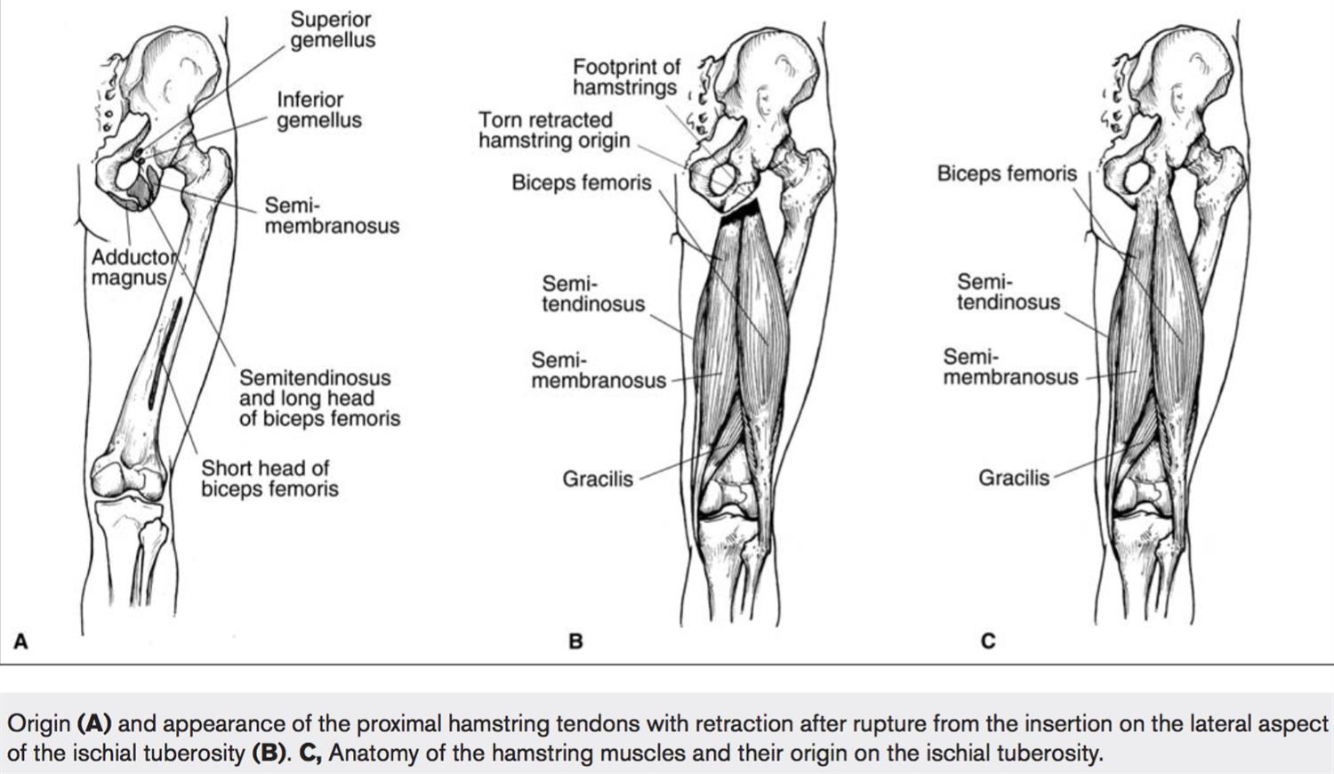 Hamstring Injuries Knee Sports Orthobullets
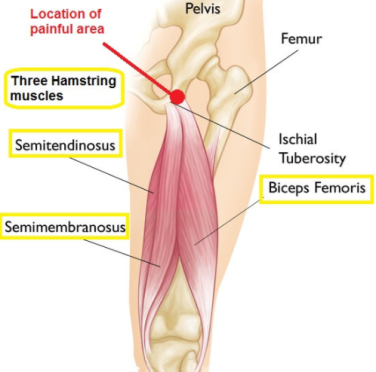 Hamstrings Injury Blaze Yoga And Pilates
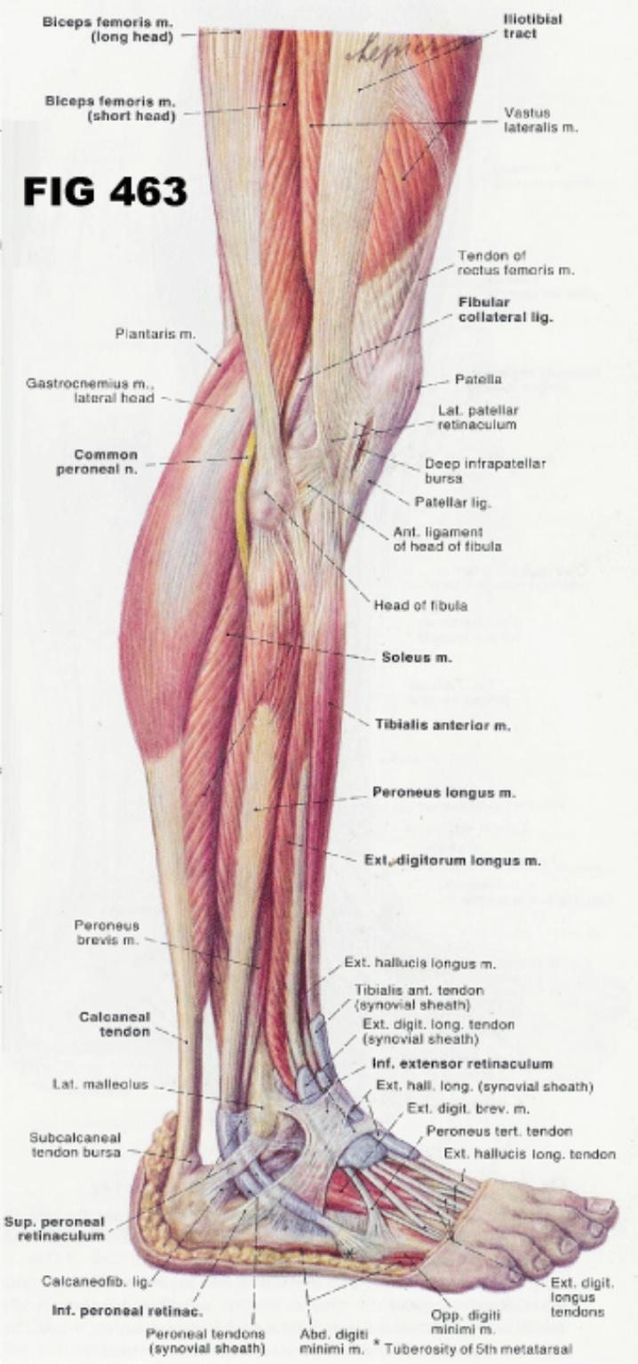 The Hamstrings And The Calves Corewalking
 Sciatic Nerve An Overview Sciencedirect Topics
 Hamstring Muscle Injuries Orthoinfo Aaos
 There Are 3 Hamstring Muscles They Are The Biceps Femoris
What Are The Best Exercises To Develop Robust Hamstrings And
 Hamstring Injury Causes Symptoms Recovery Time Treatment
 Are Your Weak Neck Muscles Making Your Hamstrings Tight
 Serious Thigh Muscle Strains Beware The Intramuscular
 Hamstring Tendon Repair Cork Ir Hamstring Injury
:max_bytes(150000):strip_icc()/the-muscles-of-the-thigh-87394652-5becaa51c9e77c00263c91d3.jpg) Anatomy Of The Hamstring Muscles
Anatomy Of The Hamstring Muscles
 A New Option For The Reconstruction Of Primary Or Recurrent
 Why Do So Many Of Us Have Tight Hamstrings Doctor Yogi


Belum ada Komentar untuk "Hamstring Muscles Anatomy"
Posting Komentar