Axillary Vein Anatomy
Axillary veins subclavian vein internal jugular vein brachiocephalic veins. In this article we shall examine the anatomy of the axilla the borders contents and any clinical correlations.
 Windsor University School Of Medicine St Kitts Ppt Video
The second part of the axillary artery gets occluded by the overlying pectoralis minor muscle when the arm was hyperabducted and brought overhead this was described by wright in 1945.

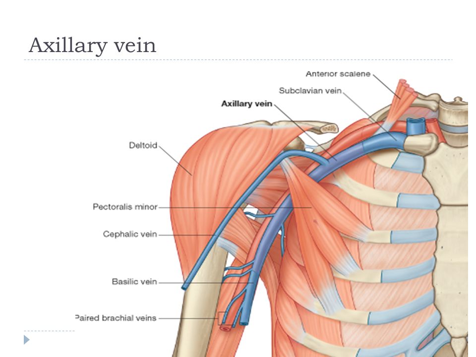
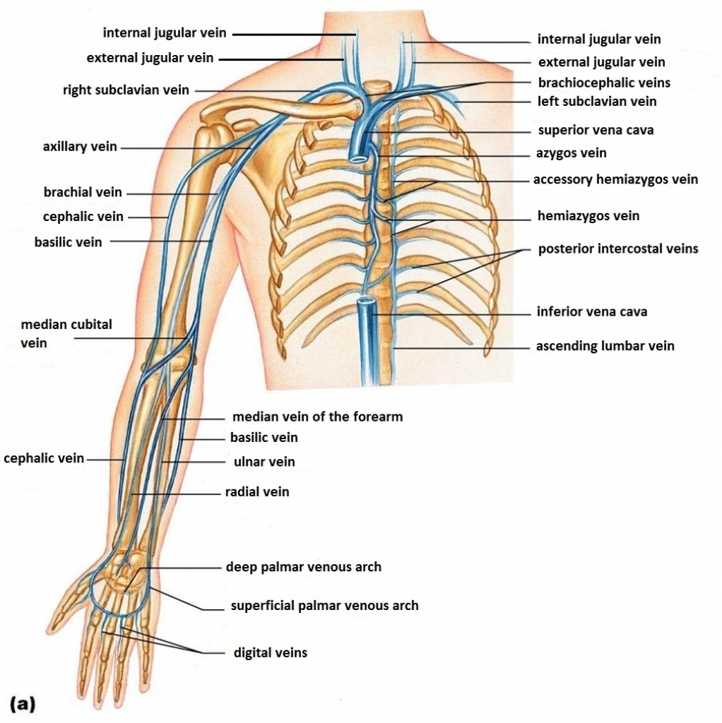
Axillary vein anatomy. The axillary vein runs along the medial side of the axillary artery. Its origin is at the lateral margin of the first rib before which it is called the subclavian artery. Upper limb veins 3d anatomy tutorial anatomyzone.
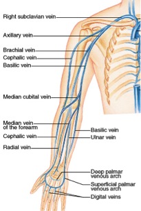
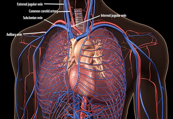
The axillary artery is the 3 rd most common site for arterial cannulation and can also be used for hemodialysis access. Here it combines with the brachial veins from the deep venous system to form the axillary vein. In human anatomy the axillary vein is a large blood vessel that conveys blood from the lateral aspect of the thorax axilla armpit and upper limb toward the heart.
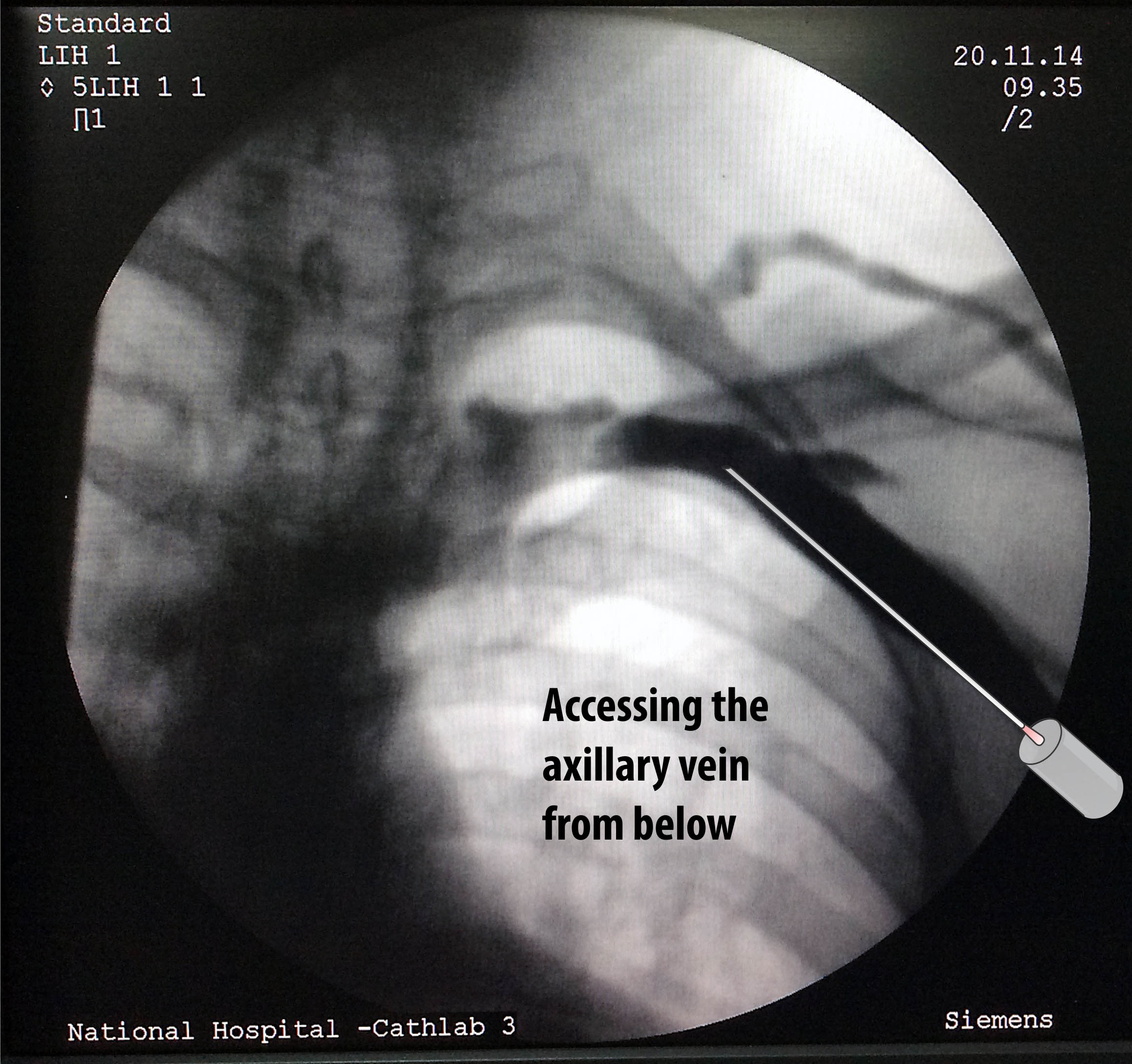
The axillary vein is formed at the inferior border of the axilla by the union of the paired brachial veins venae comitantes of the brachial artery and the basilic vein 12. There is one axillary vein on each side of the body. Keeping in common use axillary access is the preferred term as extrathoracic subclavian vein access is a mouthful.
The vein receives the axillary. From a semantics point of view this also includes the extrathoracic part of the subclavian vein. At the border of the teres major the vein moves deep into the arm.
The basilic vein originates from the dorsal venous network of the hand and ascends the medial aspect of the upper limb. In human anatomy the axillary artery is a large blood vessel that conveys oxygenated blood to the lateral aspect of the thorax the axilla armpit and the upper limb. Its origin is at the lower margin of the teres major muscle and a continuation of the brachial vein.
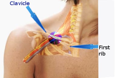
Course the axillary vein arises at the inferior border of the teres major muscle at the inferior border of the axilla 3. Axillary vein access denotes any venous access lateral to the medial border of the first rib. It begins at the lateral border of the first rib later draining into the subclavian vein.
The axilla is the name given to an area that lies underneath the glenohumeral joint at the junction of the upper limb and the thoraxit is a passageway by which neurovascular and muscular structures can enter and leave the upper limb.
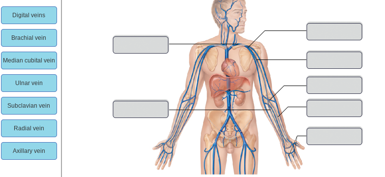 Solved Digital Veins Brachial Vein Median Cubital Vein Ul

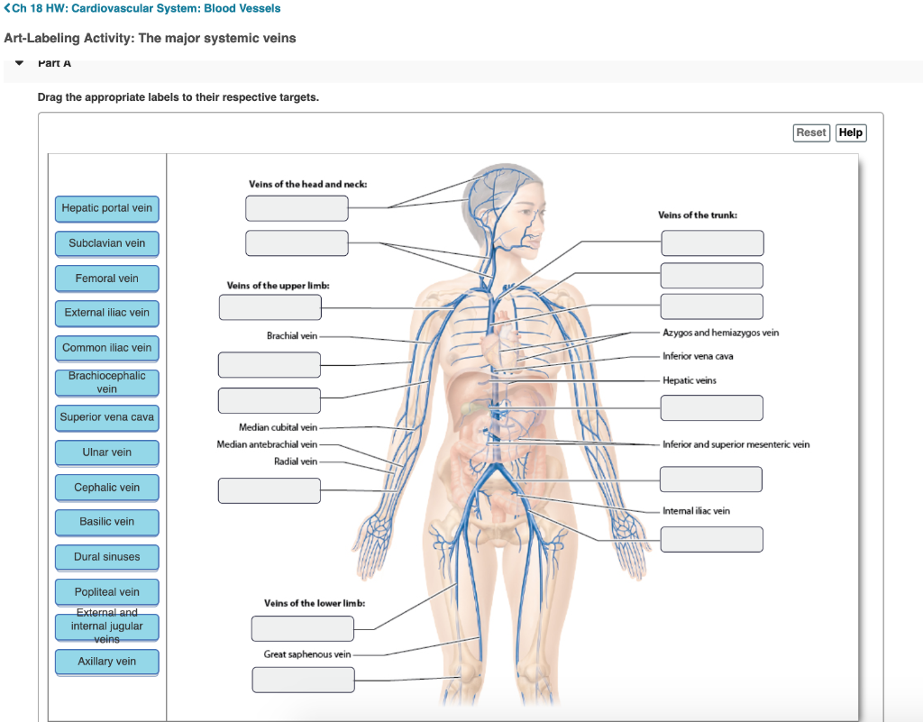 Solved Ch 18 Hw Cardiovascular System Blood Vessels Art
 Beautifully Designed Axillary Vein Art Pixels
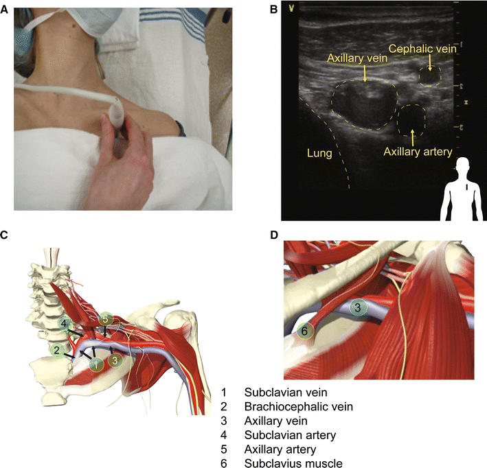 Subclavian And Axillary Vessel Anatomy A Prospective
The Axillary Vein And Its Tributaries Are Not In The Mirror
![]() Venous Contrast Injection Showing The Cephalic Vein 1
Venous Contrast Injection Showing The Cephalic Vein 1
 Axillary Vein Stock Photos Axillary Vein Stock Images Alamy

 Special Anatomical Regions Advanced Anatomy 2nd Ed
 Subclavian Vein Thrombosis Practice Essentials Anatomy

 Ultrasound Guided Subclavian Vein Cannulation The Vessel To
 Axillary Node Clearance Oncohema Key
Central Venous Access Basicmedical Key
 Cephalic Vein Superficial Vein Arm Axillary Vein Png
The Axillary Vein And Its Tributaries Are Not In The Mirror
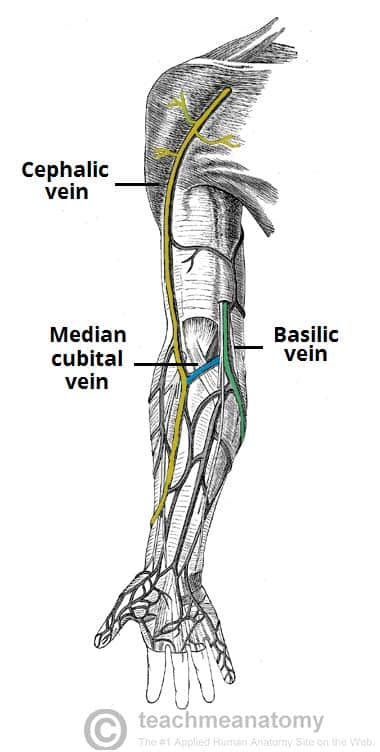 Venous Drainage Of The Upper Limb Basilic Cephalic
 Anatomy Of The Upper Extremities Gray S
![]() Cephalic Vein Anatomy And Clinical Points Kenhub
Cephalic Vein Anatomy And Clinical Points Kenhub
 Arm Dvt Normal Ultrasoundpaedia

Belum ada Komentar untuk "Axillary Vein Anatomy"
Posting Komentar