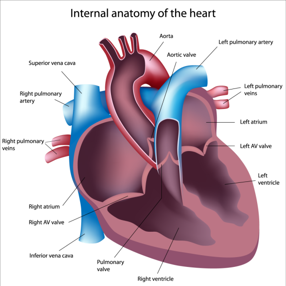
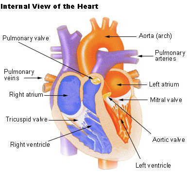
Internal Anatomy Of Heart
It is the contraction of the myocardium that pumps blood through the heart and into the major arteries. The walls and lining of the pericardial cavity are a special membrane known as the pericardium.
![]() Do Butterflies Have Brains And Hearts The Children S
Do Butterflies Have Brains And Hearts The Children S
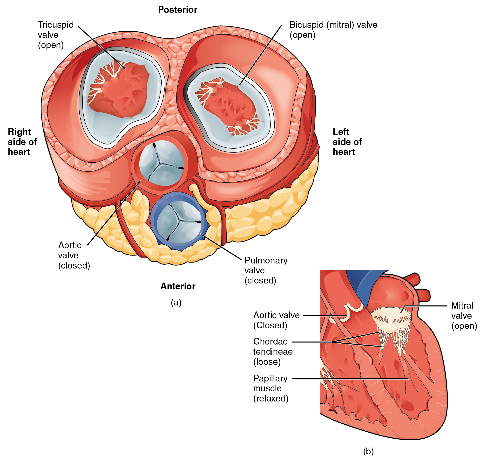
The valves between the atria and ventricles are known generically as the tricuspid right sideand the bicuspid left side va lve.
Internal anatomy of heart. Just pick an audience or yourself and itll end up in their incoming play queue. The muscle pattern is elegant and complex as the muscle cells swirl and spiral around the chambers of the heart. 0 0000 a shoutout is a way of letting people know of a game you want them to play.
Heart anatomy external the endocardium and subendocardial tissue receive oxygen and nutrients by diffusion or microvasculature directly from the chambers of the heart. Internal anatomy of the heart study guide by christinostlund includes 41 questions covering vocabulary terms and more. Anatomy of the human heart internal structures.
The heart sits within a fluid filled cavity called the pericardial cavity. The circulatory system a pdf file of the upper and lower body for printing out to use off line. The 1epicardium the 2myocardium cardiac muscle and the skip to primary navigation skip to content.
Images and pdfs. Pericardium is a type of serous membrane that produces serous fluid to lubricate the heart and prevent friction between the ever beating heart and its surrounding organs. The remainder is supplied by the coronary vasculature which is primarily embedded in the pericardial fat on the surface of the heart and supplies predominantly the epicardium.
The circulatory system lower body image with blank labels attached. The heart is situated within the chest cavity and surrounded by a fluid filled sac called the pericardium. The anatomy of the heart.
Internal structure of the heart. The heart an image of the heart with blank labels attached. They form a figure 8 pattern around the atria and around the bases of the great vessels.
It is divided by a partition or septum into two halves and the halves are in turn divided into four chambers. The valves at the openings that lead to the pulmonary trunk and aorta are known generically as the pulmonary and the aortic valve. The circulatory system upper body image with blank labels attached.
The heart ventricular walls consist of three layers. Quizlet flashcards activities and games help you improve your grades. This amazing muscle produces electrical impulses that cause the heart to contract.
 Male Internal Anatomy Of Heart
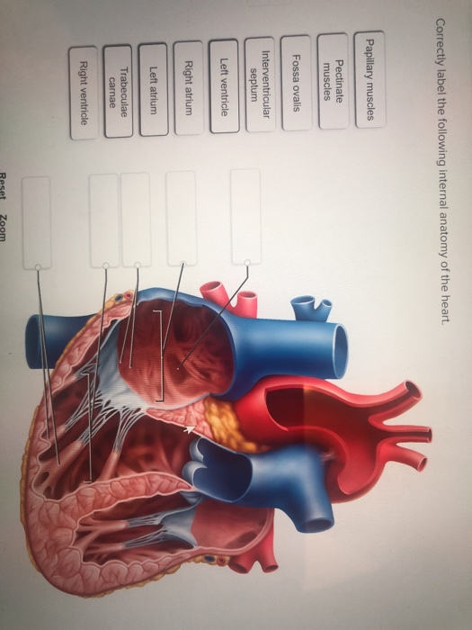 Solved Correctly Label The Following Internal Anatomy Of
 Internal Anatomy Of Heart Left Side 1 Diagram Quizlet

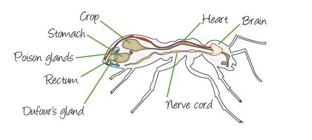
Internal Ant Anatomy Brain Heart Nerve Cord Crop
 Is Pulmonary Arterial Hypertension A Heart Disease Or Lung
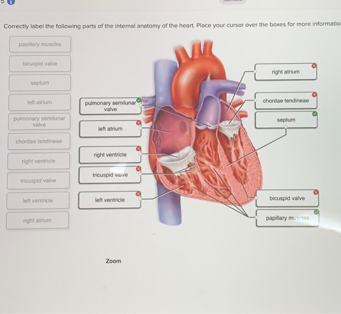 Solved Correctly Label The Following Parts Of The Interna
 Human Heart Diagram And Anatomy Of The Heart Heart

 Chapter 19 The Heart Circulatory System Cardiovascular
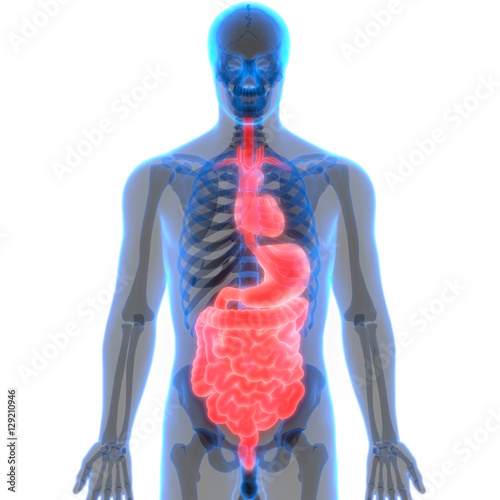
 Abdomen Anatomy Definition Function Muscles Biology
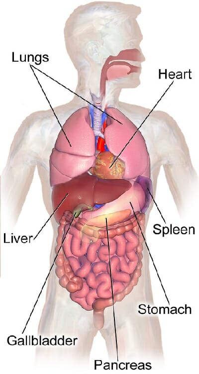
 Human Body Organs Anatomy Heart With Digestive System
 Free Anatomy Quiz Anatomy Of The Heart Quiz 1
![]() A Transparent Human Heart With Internal Anatomy Visible
A Transparent Human Heart With Internal Anatomy Visible

 Amazon Com Vision Scientific Vsa471 Jumbo Human Model 3
 Structure And Function Of The Heart Course Hero
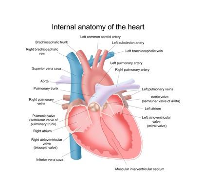 Illustration Of The Internal Anatomy Of The Heart Science
 Seer Training Structure Of The Heart
 Heart Anatomy Internal Medical Art Library
 Heart Anatomy Anatomy And Physiology Ii
 19 1 Heart Anatomy Anatomy And Physiology
 Organs Anatomy Stock Photos Organs Anatomy Stock Images
 Heart Anatomy Chambers Valves And Vessels Anatomy
 Label The Heart Purposegames
 Human Heart Anatomy
 Internal Anatomy Of The Heart Flashcards Quizlet
Belum ada Komentar untuk "Internal Anatomy Of Heart"
Posting Komentar