Leg Venous Anatomy
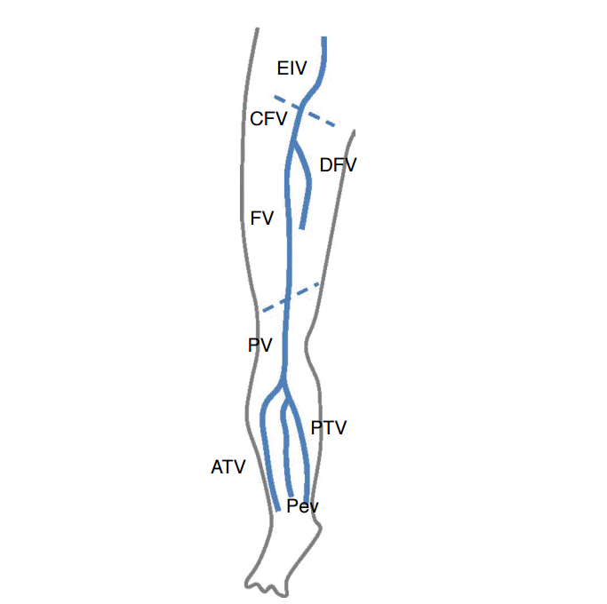
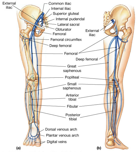
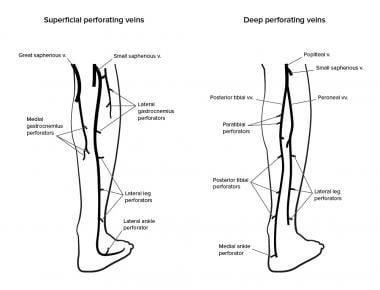
The plantar venous arch sends its blood into the leg through the medial and lateral plantar veins into the posterior tibial vein which ascends along the leg posterior to the tibia. Superficial veins of the lateral leg and thigh form the lateral venous system.
 Diagram Showing The Venous Anatomy Of The Leg
The plantar and the dorsal veins.

Leg venous anatomy. Each individual hands on training case is accompanied by image window specific expert instruction and probe positioning guidance. Deep veins of the foot form two divisions. On the plantar aspect of the foot medial and lateral plantar veins arise.
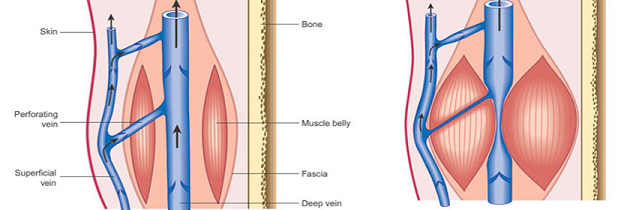
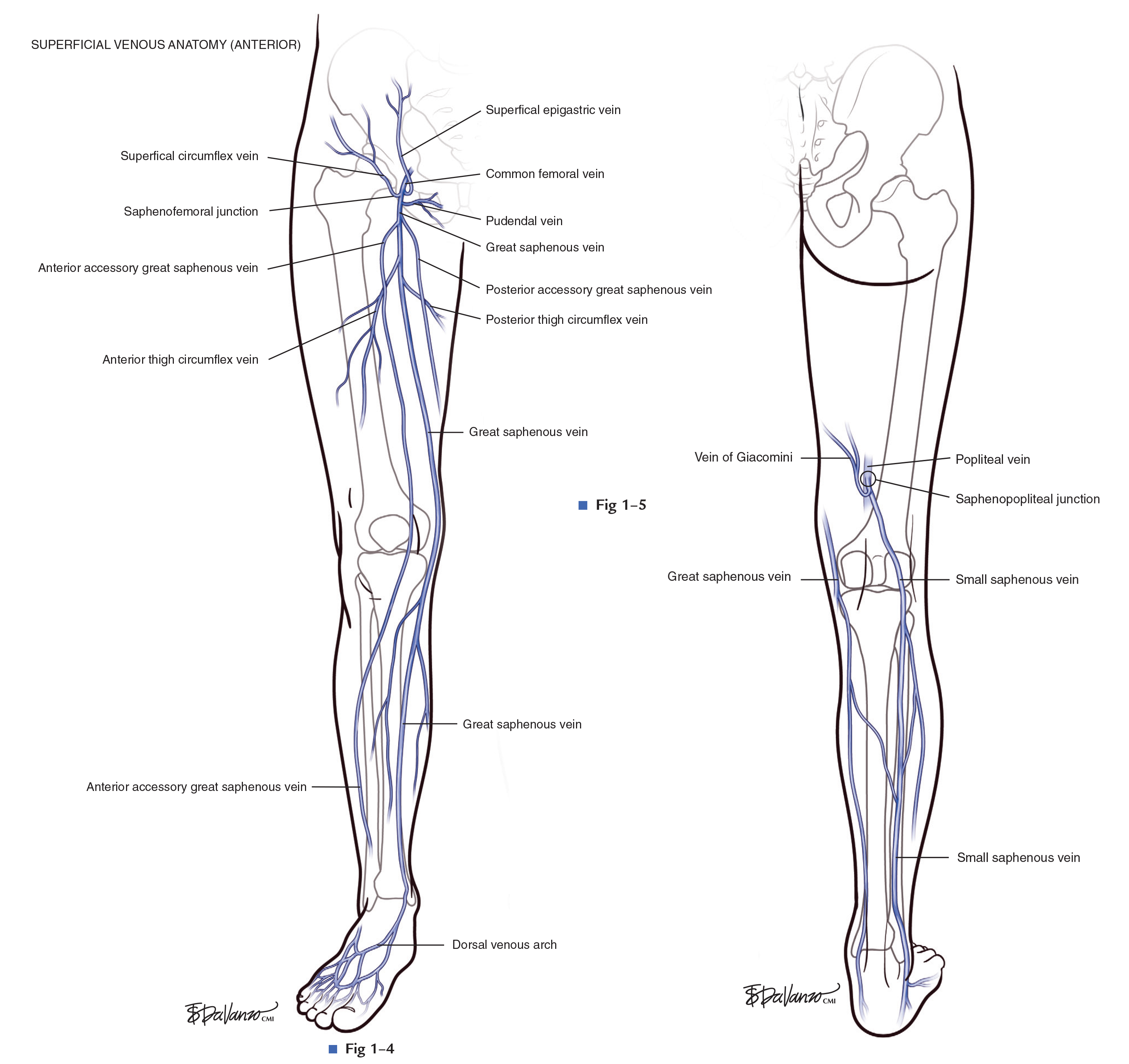
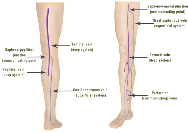
The lateral venous system is drained through multiple small tributaries into the gsv and ssv. And the perforating veins that penetrate the muscular fascia and connect the superficial and deep veins. Some veins from the arch penetrate deep into the leg forming the anterior tibial vein.
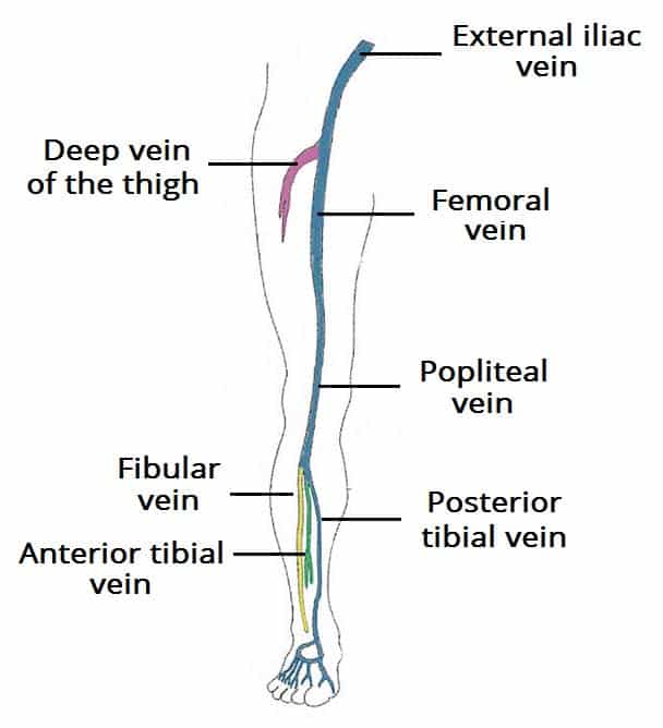
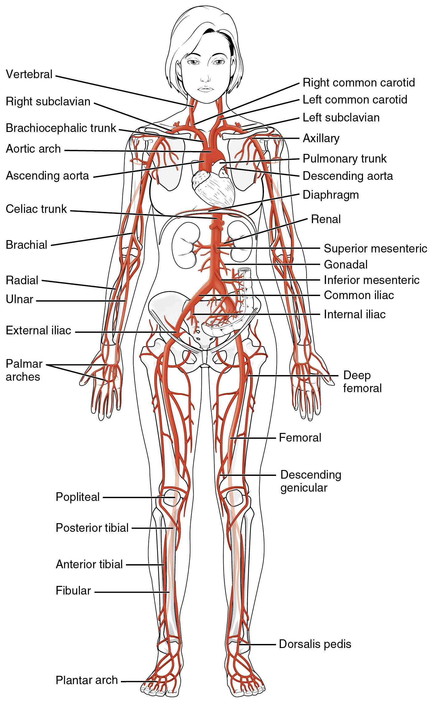
This article will discuss the anatomy and tributaries of the veins of the lower limb in detail followed by any related clinical notes. The venous system of the lower extremities includes the deep veins which lie beneath the muscular fascia and drain the lower extremity muscles. Anterior tibial vein which receives blood from the dorsal venous arch.
The superficial veins which are above the deep fascia and drain the cutaneous microcirculation. The sural nerve courses along the ssv in the distal calf. These veins combine to form the posterior tibial and fibular veins.
The main venous structure of the foot is the dorsal venous arch which mostly drains into the superficial veins. The anterior tibial vein forms a small network anterior to the tibia and collects blood from the tissues of the shin. Posterior tibial vein and fibular vein also known as the peroneal vein which form from the medial and lateral plantar veins.
Anatomy physiology module provides a broad spectrum of adult male adult female and pediatric normal anatomy cases with varying body morphologies to maximize training efficacy. Both types of veins contain venous valves to prevent reflux of blood distally but they are more numerous in the deep veins. They also contain tributaries other veins which drain into them.
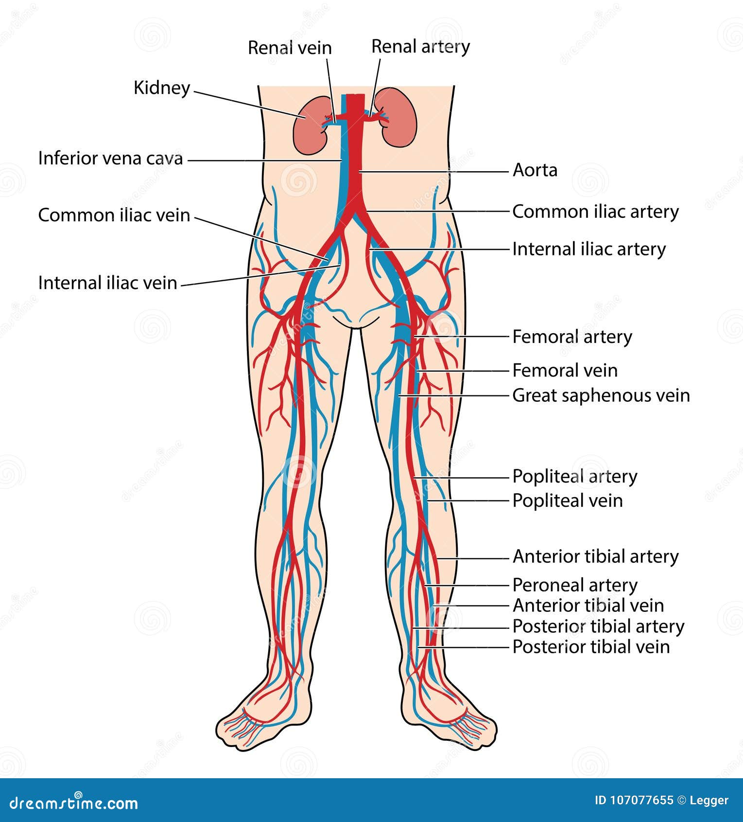
Veins of the lower limb. There are three main deep veins in the lower leg.
 Ultrasonography
 Vein Services Biltmore Cardiology
 Intro To Venous Reflux
 Leg Knee Anatomy
 Venous Drainage Of The Lower Limb Teachmeanatomy
 Emdocs Net Emergency Medicine Educationcore Em
 Vein Wikipedia
 Venous Anatomy Milwaukie Vein Center
 Assessment And Management Of Patients With Varicose Veins
 Leg Vein Anatomy Stock Illustrations Images Vectors
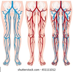
 20 5 Circulatory Pathways Anatomy And Physiology
 Imbus Home
 Blood Vessels Of The Lower Limbs Course Hero
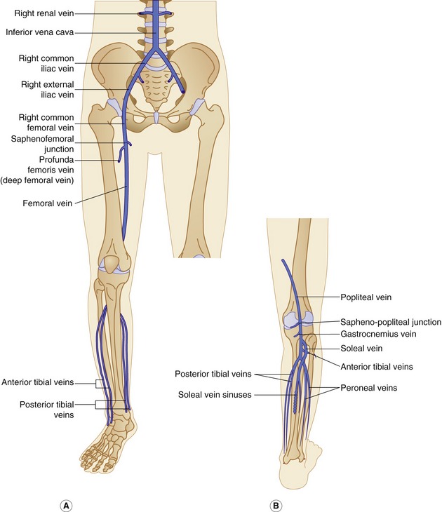
 Venous Drainage Of The Lower Extremity Anatomy
 Pediagenosis
 Vein Anatomy And Function Vein Human Leg
 Figure 4 From The Hemodynamics And Diagnosis Of Venous
 Leg Vein Anatomy Los Angeles Arterial Leg Information
 Lower Extremity Veins Radiology Key
 Blood Vessels Of The Lower Body Stock Vector Illustration
 Anatomy Of The Lower Limb Venous System And Assessment Of
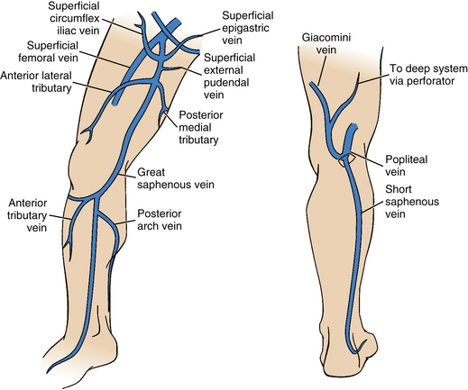
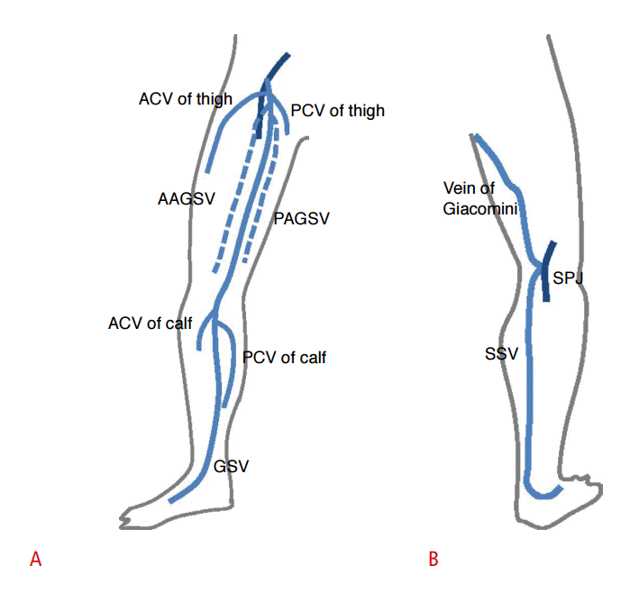
 Anatomy Of Gsv And Ssv With Common Variants Of Ssv Gsv
 Ultrasonography
 Venous Insufficiency Background Anatomy Pathophysiology
 Femoral Vein Wikipedia
 Lower Leg Venous Anatomy Admirable Upper Extremity Anatomy
 Venous Reflux Study With Ultrasound The Vein Institute Of
 Leg Venous Anatomy Physiology Module Sonosim
 Chronic Venous Insufficiency Advanced Vein Care
 Leg Vein Anatomy 101
 Imagenes Fotos De Stock Y Vectores Sobre Veins Leg Anatomy
 Image Result For Lower Extremity Venous Anatomy Arteries
Leg Vein Anatomy Veinspecialistsofarizona Com
 Venous Anatomy And Physiology Venous And Lymphatic


Belum ada Komentar untuk "Leg Venous Anatomy"
Posting Komentar