Skull X Ray Anatomy
Heres a quick lecture on the basic radiology of the skull which you will be held responsible for on your midterm. Cranial bones 8 the eight bones of the cranium are divided into the.
 X Ray Image Or Roentgen Of Human Skull Close Up Head Xray Scan
We would like to show you a description here but the site wont allow us.
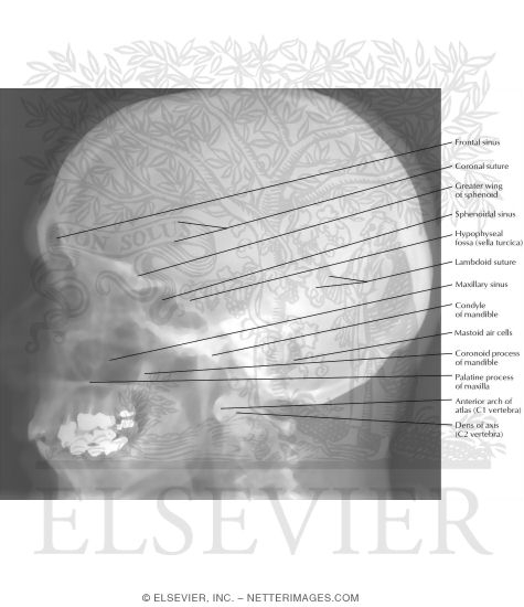
Skull x ray anatomy. Petrosal bone of temporal bone. It consists of 8 cranial bones and 14 facial bones see our article on radiographic positioning of the face and mandible. The anatomy of the skull is very complex and specific attention to detail is required of the technologist.
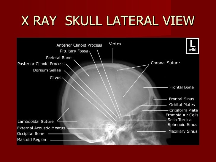
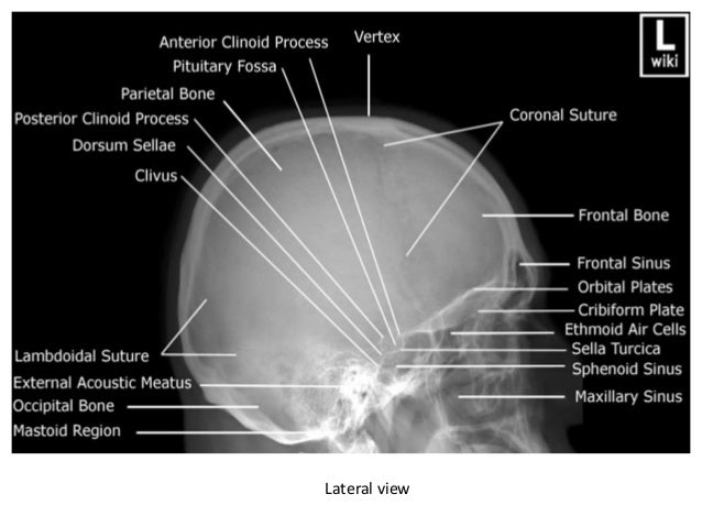
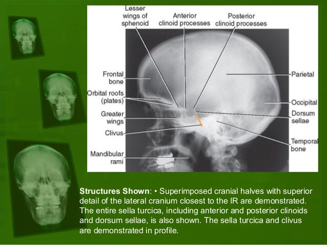
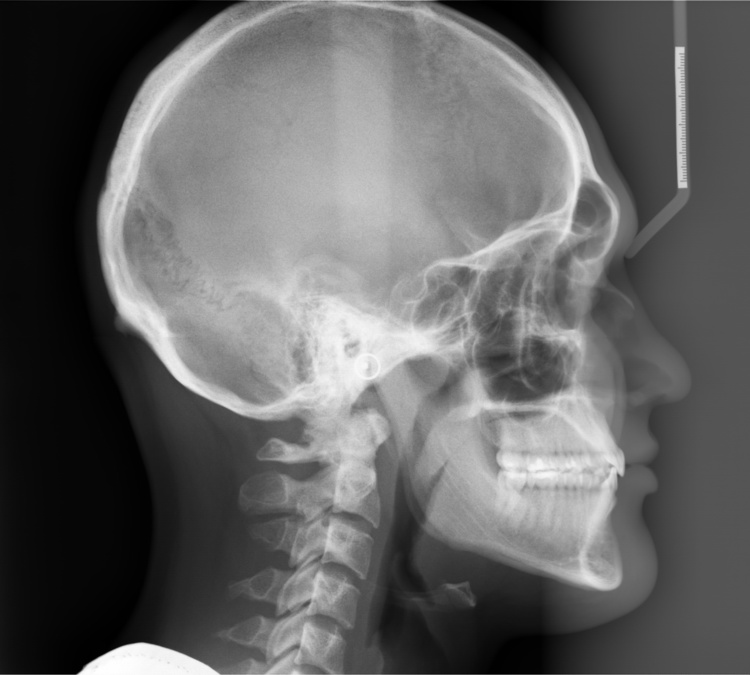
Superior margin of. Skull x ray lateral view this is an x ray image of the skull taken from a lateral view showing the skull from the side. The back of the cranium consists of the occipital and right and left parietal bones.
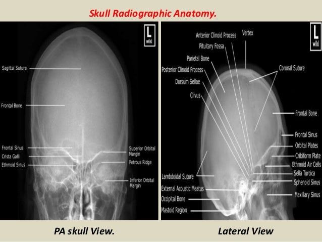
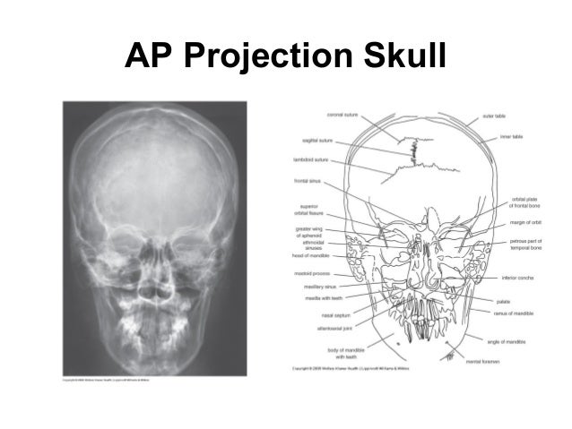
Patient position the back of patients head is placed against the image detector. Close collimation also reduces the radiation dose to the patient and the technologist. Skull x rays show the course of vessels which indent the inner table these vascular indentations branch and taper whereas fractures do not usually branch or taper normal skull ap.
Skull ap with 3d. Chest x ray cxr. X ray examination of the skull that are routinely done in radiology department and includes a brief anatomy.
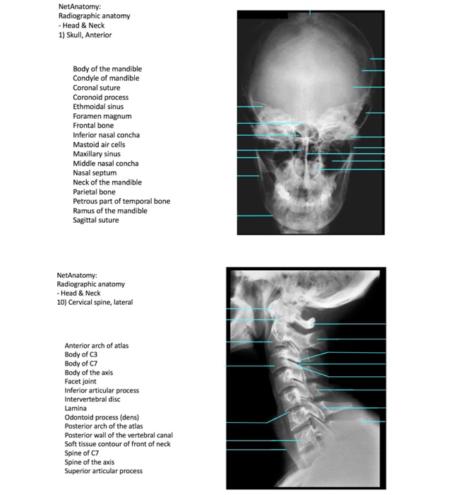
Radiographic anatomy skull as with other body parts radiography of the skull requires a good understanding of all related anatomy. The skull is a solid bony structure that encloses and protects the brain and other components of the central nervous system. The skull or bony skeleton of the head rests on the superior end of the vertebral column and is divided into two main sets of bonesthe 8 cranial bones and the 14 facial bones.
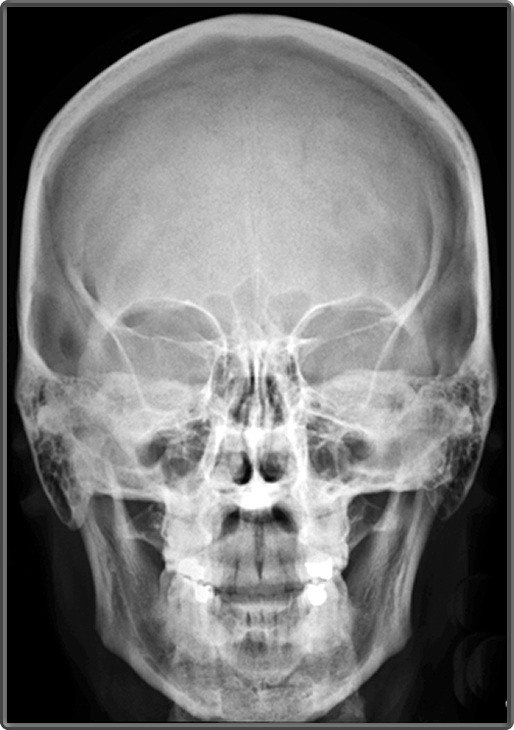
Radiographic anatomy of the cervical spine full. The skull ap view is a nonangled ap radiograph of the skull. Close collimation of the x ray field reduces the amount of tissue irradiated reducing the amount of scatter pro duced and increasing contrast.
X ray beam the amount of tissue irradiated and the type and thick ness of the tissue. This view provides an overview of the entire skull rather than attempting to highlight any one region.
 Film X Ray Radiograph Show Anatomy Stock Photo Edit Now
 Radiology Quiz 8301 Radiopaedia Orgviewing Playlist Uq
 Female Occipital Bone Skull Anatomy Isolated On White Art Print Poster
![]() Student Study Guides Skull Anatomy
Student Study Guides Skull Anatomy
 Film X Ray Radiograph Show Human Anatomy Of Skull Bone And
 Presentation1 Radiological Imaging Of Fractures
 Emergency X Ray Interpretation Part 7 Skull And Face
 Normal Skull X Ray Stock Image C039 4286 Science
 Frontal Radiograph Of Skull Medical Radiography Anatomy
 Plain X Ray Skull
 Head Neck Rs X Rays

 Brain Anatomy Medical Head Skull Digital 3 D X Ray Xray
 Positioning And Radiographic Anatomy Of The Skull
 Skull X Ray Stock Image C003 4553 Science Photo Library
 Human Brain Shirt Vintage Anatomy Skull X Ray Gift
 Head Skull X Ray Buy This Stock Photo And Explore Similar
 Anatomy Xray Picture Human Skull Stock Photo Edit Now
 Cephalometric X Ray Dental Diagnostic Imaging Center
 X Ray Image Of Skull Human
Skull Lateral Radiograph
 Radiographic Anatomy Of Facial Bones Pdf

 The Canine Head And Skull Ct Atlas Of Veterinary Clinical
 Linear Skull Fracture In The Simple X Ray And 3 Dimensional
 A Gross Anatomy And B Dorsoventral Radiographic View Of
Belum ada Komentar untuk "Skull X Ray Anatomy"
Posting Komentar