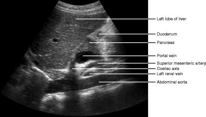
Pancreas Ultrasound Anatomy
Pancreatic ducts for more information. Give the patient an oral waterload 2 3 glasses and scan them erect.
 Startradiology
Learn the conditions that affect the pancreas as well as its function and location in the body.

Pancreas ultrasound anatomy. Ultrasound of the pancreas. Imaging features of normal pancreas grafts us imaging. 189 layers of adrenal gland.
The anterior and posterior superior pancreaticoduodenal veins drain directly into the portal vein. Improved visualisation of the pancreas after a water load. As would be expected a transplanted pancreas has the same imaging features as.
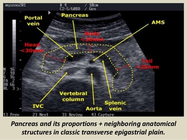
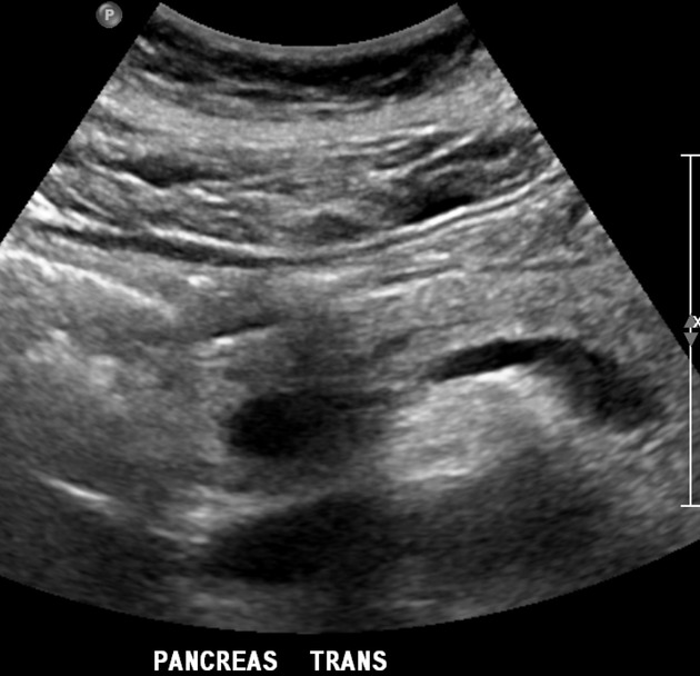
Ultrasound examination of the pancreas in the transversal direction. The water is used as a window to look through when it is in the stomach and duodenum. The left adrenal gland is frequently crescent shaped.
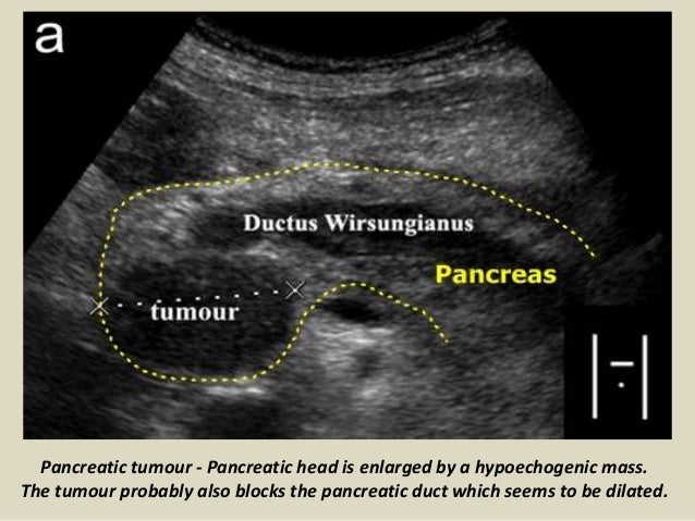
Webmds pancreas anatomy page provides a detailed image definition and information about the pancreas. Pancreatic ultrasound can be used to assess for pancreatic malignancy pancreatitis and its complications as well as for other pancreatic pathology. Preparation fast the patient to reduce interference from overlying bowel gas which may otherw.
The head of the pancreas is drained by the two anterior and posterior inferior pancreaticoduodenal veins which empty into the superior mesenteric vein. The pancreas before water and mouse over after 2 glasses of water. Mr imaging evaluation of pancreas grafts is.
The pancreas is an abdominal glandular organ with an digestive exocrine and hormonal endocrine function. Pancreatic juice is secreted into a branching system of pancreatic ducts that extend throughout the gland. In this article we shall look at the basic anatomy of the pancreas.
Because of its position the pancreas is not always fully visible in any plane fig. The left adrenal gland often extends relatively far downward toward the renal hilum. In the majority of individuals the main pancreatic duct empties into the second part of duodenum at the ampulla of vater.
Angulating the echotransducer andor moving it in the craniocaudal direction will allow for evaluation of as much of the pancreatic parenchyma as possible. The relatively complex vascular anatomy of a pancreas transplant is usually best displayed.
 Ultrasound Training Pancreas
Chapter 8 Ultrasound Of The Pancreas Surgical And
 Ultrasound Of Pancrease In Radiology
 Startradiology
 Imaging Of The Pancreas Radiology Key
 Diagnostics Free Full Text The Role Of Transabdominal
 Computed Tomography Ct Scan Of The Pancreas Johns
 Ultrasound Of The Pancreas What Normal Looks Like
Endoscopic Ultrasound Of Pancreatic Lesions Chong The
 Startradiology
 Transabdominal Ultrasonography Of The Pancreas Basic And
 Imaging The Olive Laboratory
 Imaging Of The Pancreatic Duct By Linear Endoscopic Ultrasound
 Imaging Of The Pancreas Radiology Key
 Pancreatic Ultrasound Radiology Reference Article
 Small Animal Abdominal Ultrasonography Today S Veterinary
 Chapter 8 Ultrasound Of The Pancreas Surgical And
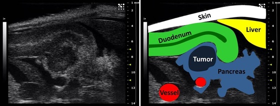
 Pancreatic Neuroendocrine Tumor Radiology Case
 Abdominal Ultrasound Registry Review
 Ultrasound Of The Pancreas
 Pancreas And Spleen Springerlink
 Chapter 8 Ultrasound Of The Pancreas Surgical And
 Imaging Of The Pancreatic Duct By Linear Endoscopic
 Pancreas Anatomy Overview Gross Anatomy Microscopic Anatomy
 Abdominal Ultrasound Registry Review
 Ultrasound Of The Pancreas What Normal Looks Like
 Roadmap Developed For Role Of Focused Ultrasound In Treating
 Pdf Don T Forget About The Pancreas Useful Tips On Ultrasound
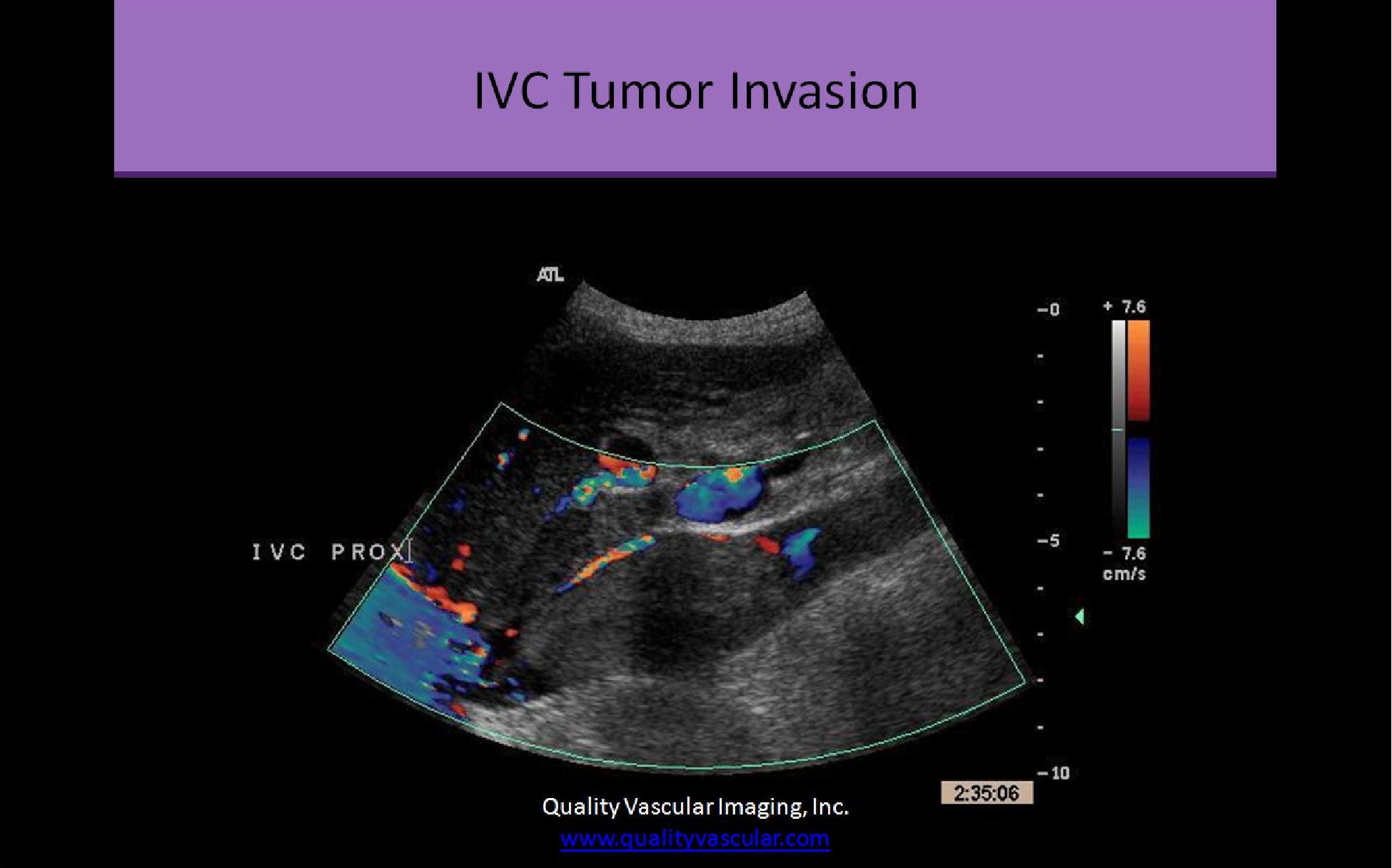
 Midline Abdomen Ultrasound Sonography Vascular Ultrasound
![]() Startradiology
Startradiology
 Ultrasound Of The Pancreas What Normal Looks Like
 Ultrasound Of The Pancreas What Normal Looks Like
 Ultrasound Of Pancrease In Radiology
 Small Animal Abdominal Ultrasonography Today S Veterinary


Belum ada Komentar untuk "Pancreas Ultrasound Anatomy"
Posting Komentar