Anatomy Of The Knee Diagram
The knee joins the thigh bone femur to the shin bone tibia. The anatomy of the knee consists of bones muscles nerves cartilages tendons and ligaments.
 The Knee Anatomy Injuries Treatment And Rehabilitation
Participation in sports and recreational activities are risk factors for knee injury.

Anatomy of the knee diagram. The largest joint in the body the knee moves like a hinge allowing you to sit squat walk or jump. The knee is the largest and most complex joint in the body. All these parts combine and work together.
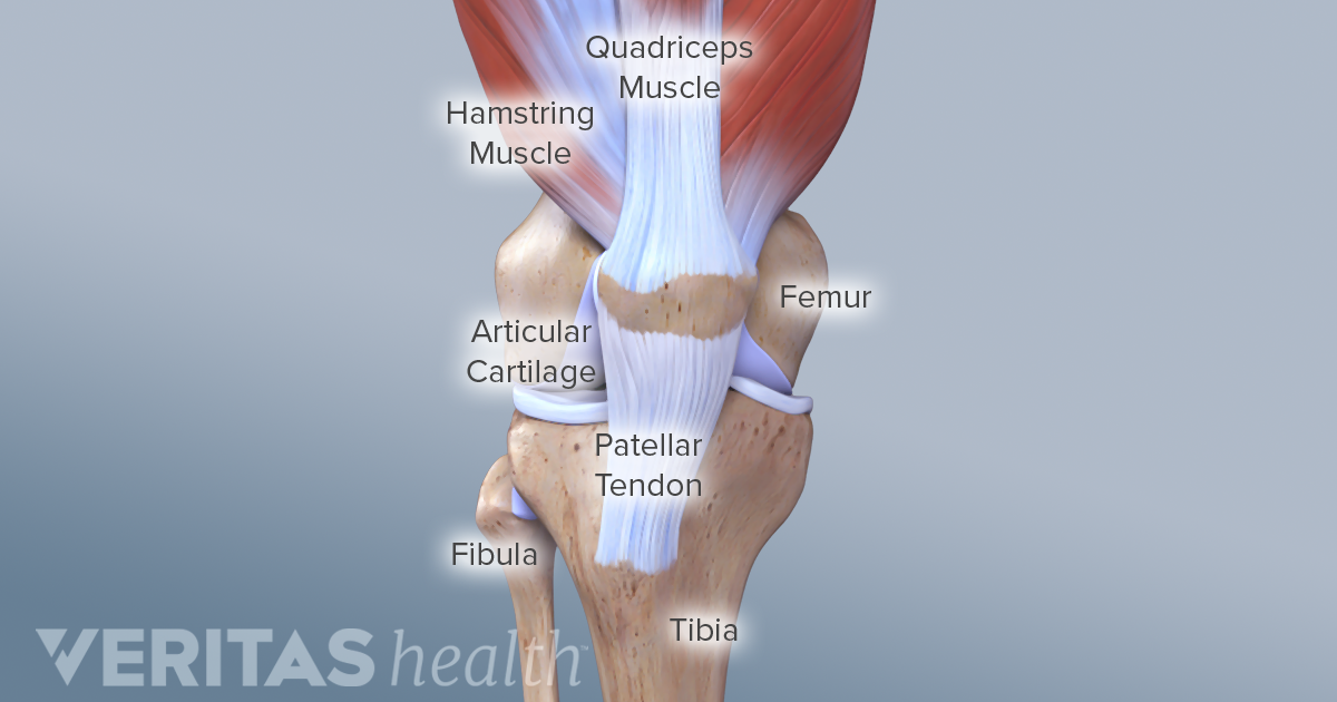
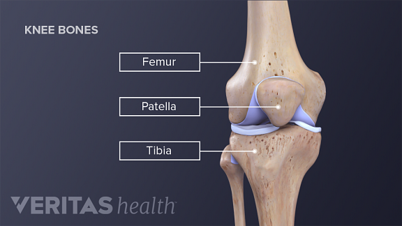
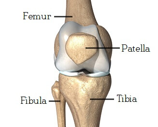
The knee consists of three bones. The kneecap glides in a groove in the thighbone and adds leverage to the thigh muscles which are used to extend the leg. The knee is the joint where the bones of the lower and upper legs meet.
Femur the upper leg bone or thigh bone. Knee joint is one of the most important hinge joints of our body. The thigh bone and shine bone come together at the knee joint and move on one another when bending or straightening the leg.
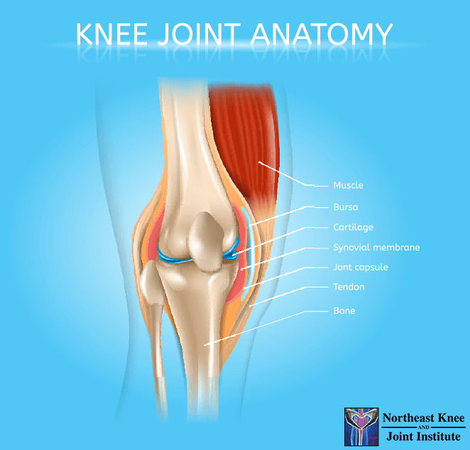
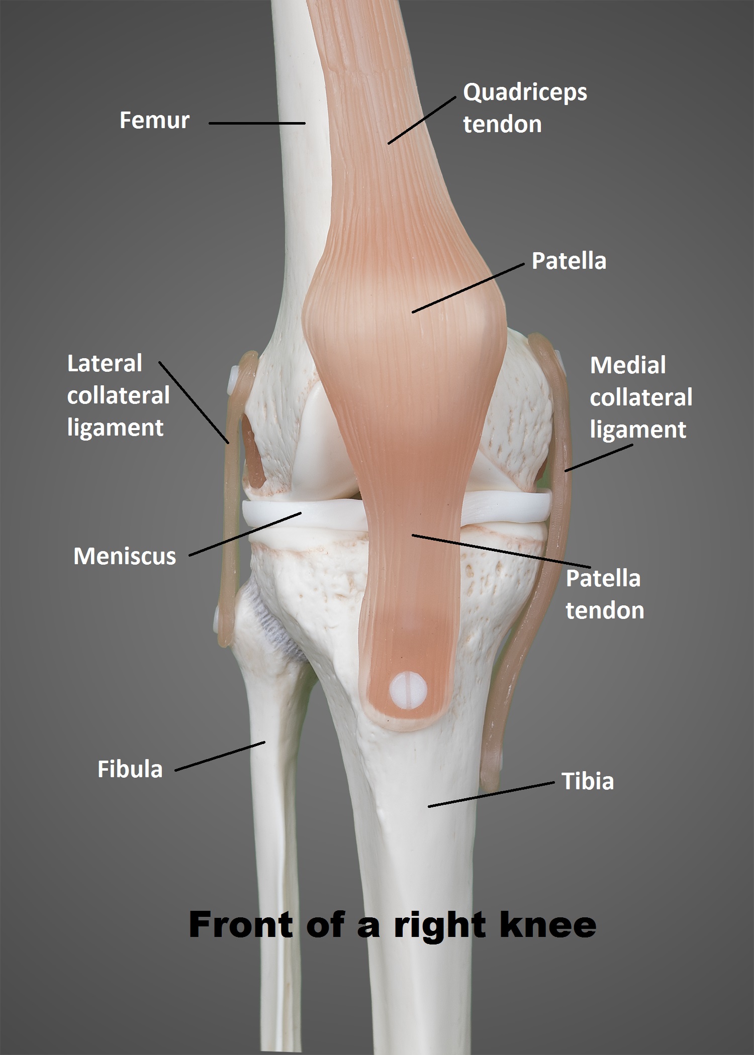
Muscles tendons and ligaments connect the knee bones. The knee is the meeting point of the femur thigh bone in the upper leg and the tibia shinbone in the lower leg. There are two main joints in the knee.
They are they soft tissues found at the end of muscles which link the muscle to bone. Another bone the patella kneecap is at the center of the knee. The knee joins together the thigh bone shin bone fibula on the outer side of the shin and kneecap.
The knee is one of the largest and most complex joints in the body. Labeled diagram of the knee joint. Fast facts on knee anatomy.
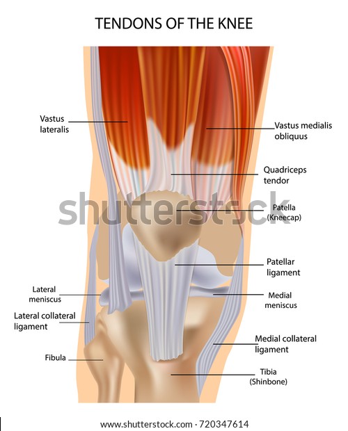
Tendons at the knee. The knee is a synovial joint meaning it contains a fluid filled capsule. 1 the tibiofemoral joint where the tibia meet the femur 2 the patellofemoral joint where the kneecap or patella meets the femur.
The knee cap actually sits inside the patellar tendon. Tendons are often overlooked as part of knee joint anatomy. The smaller bone that runs alongside the tibia fibula and the kneecap patella are the other bones that make the knee joint.
Tibia the bone at the front of the lower leg or shin bone. Tendons connect the knee bones to the leg muscles that move the knee joint. The fibula calf bone the other bone in the lower leg is connected to the joint but is not directly affected by the hinge joint action.
The main tendon found at the knee is the patellar tendon which links the quads muscles to the shin bone. The knee joint is a synovial joint which connects the femur thigh bone the longest bone in the body to the tibia shin bone. Its complexity and its efficiency is the best example of gods creation.

 Knee Anatomy Muscles Tendons Muscle Structure Stock Vector
/188058334-crop-56aae7425f9b58b7d0091480.jpg) What Is Causing Your Knee Pain
What Is Causing Your Knee Pain
 Knee Anatomy

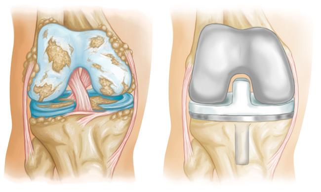 Knee Replacement Implants Orthoinfo Aaos
 Total Joint Anatomy Lakeshore Orthopaedics
 Knee Posterior Approach Approaches Orthobullets
 Figure Anatomy Of The Right Knee Download Scientific Diagram
 Knee Anatomy
 What Are The Parts Of The Knee Joint Systems4knees
 6 Types Of Arthritis That Affect The Knee
 The Knee Joint Articulations Movements Injuries
 Hyaluronic Acid Injections Northeast Knee Joint Institute
 Knee Joint Picture Image On Medicinenet Com
 Anatomy Of The Knee Joint Download Scientific Diagram
 Knee Injuries For Parents Nemours Kidshealth
 Anatomy Of Human Knee Joint See Online Version For Colours
 Anatomy Of The Knee Central Coast Orthopedic Medical Group
 Understanding The Anatomy Of The Knee Bodyheal
 Knee Joint Anatomy Motion Knee Pain Explained
Redding Hospital Knee Anatomy
 The Knee Ut Health San Antonio
Collateral Ligament Injuries Orthoinfo Aaos
Belum ada Komentar untuk "Anatomy Of The Knee Diagram"
Posting Komentar