Anatomy X Ray
The bodily structure of a plant or an animal or of any of its parts. The most frequently used imaging modalities are radiography x ray computed tomography ct and magnetic resonance imaging mrix ray and ct require the use of ionizing radiation while mri uses a magnetic field to detect body protons.

X ray anatomy what others are saying dentistry lectures for mfdsmjdfnbdeore.

Anatomy x ray. Ct scan of the scapula example 3. In a stress x ray the physician will apply pressure on the affected ankle while an x ray is being taken. Ct scan of the scapula example 2.
The radiation is created when an electric current is generated from a high voltage generator causing electrons to boil off from the cathode end of an x ray tube assembly. Ct scan of the scapula example 4. In fact some important structures such as the phrenic nerve are not visible at all.
Radiographic anatomy of facial bones and mandible with radiological abnormalities of the skull and facial bones. It gathers several non invasive methods for visualizing the inner body structures. Ct scan of the scapula example 1.
X ray of forearm and elbow extension. The science of the shape and structure of organisms and their. X ray of coronoid process.
Test yourself chest x ray quiz 1. X ray of hands and wrists. X ray anatomy synonyms x ray anatomy pronunciation x ray anatomy translation english dictionary definition of x ray anatomy.
Medical imaging is where your human anatomy knowledge meets clinical practice. X ray of carpal bones. X ray of the shoulder.
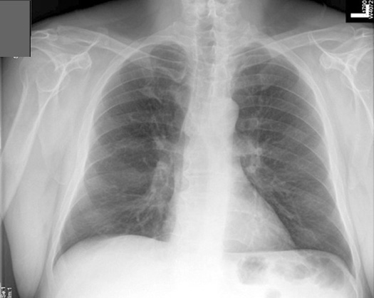
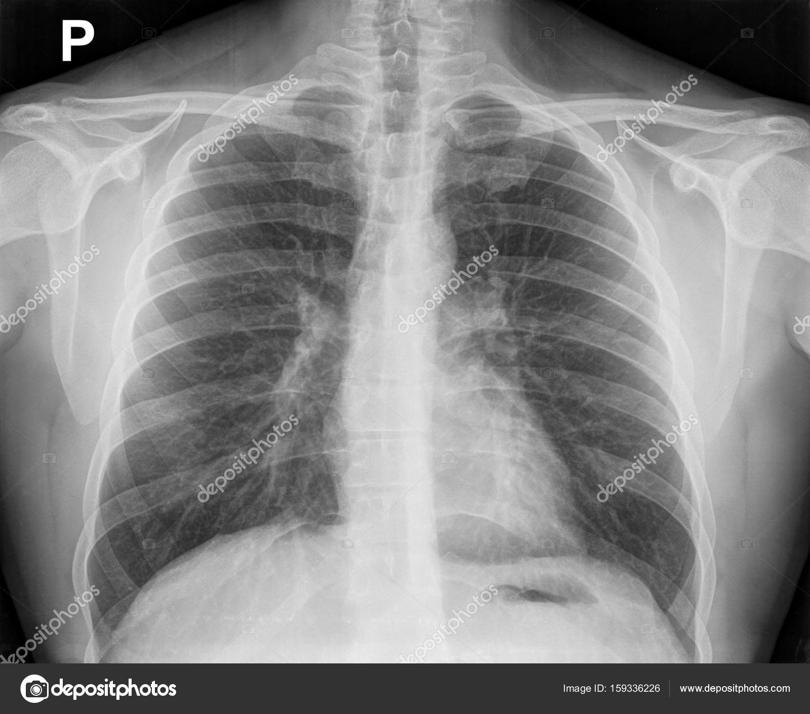
X ray of the chest also known as a chest radiograph is a commonly used imaging study and is the most frequently performed imaging study in the united states. X ray of forearm and elbow flexion. Mri scan of the shoulder.
The x ray is the most commonly requested radiographic examination because of its availability. As they are used in medicine x rays are emitted from an x ray machine and directed toward a specially treated metallic plate placed behind the patients body. Radiology and medical imaging tutorials for uk medical students.
The x ray is a form of high energy electromagnetic radiation with a short wavelength capable of penetrating solids and ionizing gases. Plain film x ray is the most common diagnostic radiological modality used in hospitals today. Other anatomical structures such as the pleura only become clearly visible when abnormal.
It is almost always the first imaging study ordered to evaluate for pathologies of the thorax although further diagnostic imaging laboratory tests and additional physical examinations may be necessary to help confirm the diagnosis. This test will be able to show any crack or break in the bones in the ankle 4 9. Chest x ray anatomy many structures of the chest are readily visible on a chest x ray but others are difficult to see.
 Details About Framed Print Funny X Ray Love Heart Hands Picture Poster Medical Anatomy Art
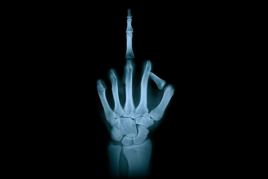 Hd Wallpaper Hand Middle Finger X Ray Radiation Finger
 Anatomy Of Human Urogenital Organs In X Ray View Canvas Print
 Manus X Ray Anatomy Radiology Radiographic Stock Photo Edit
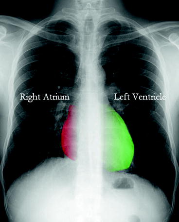
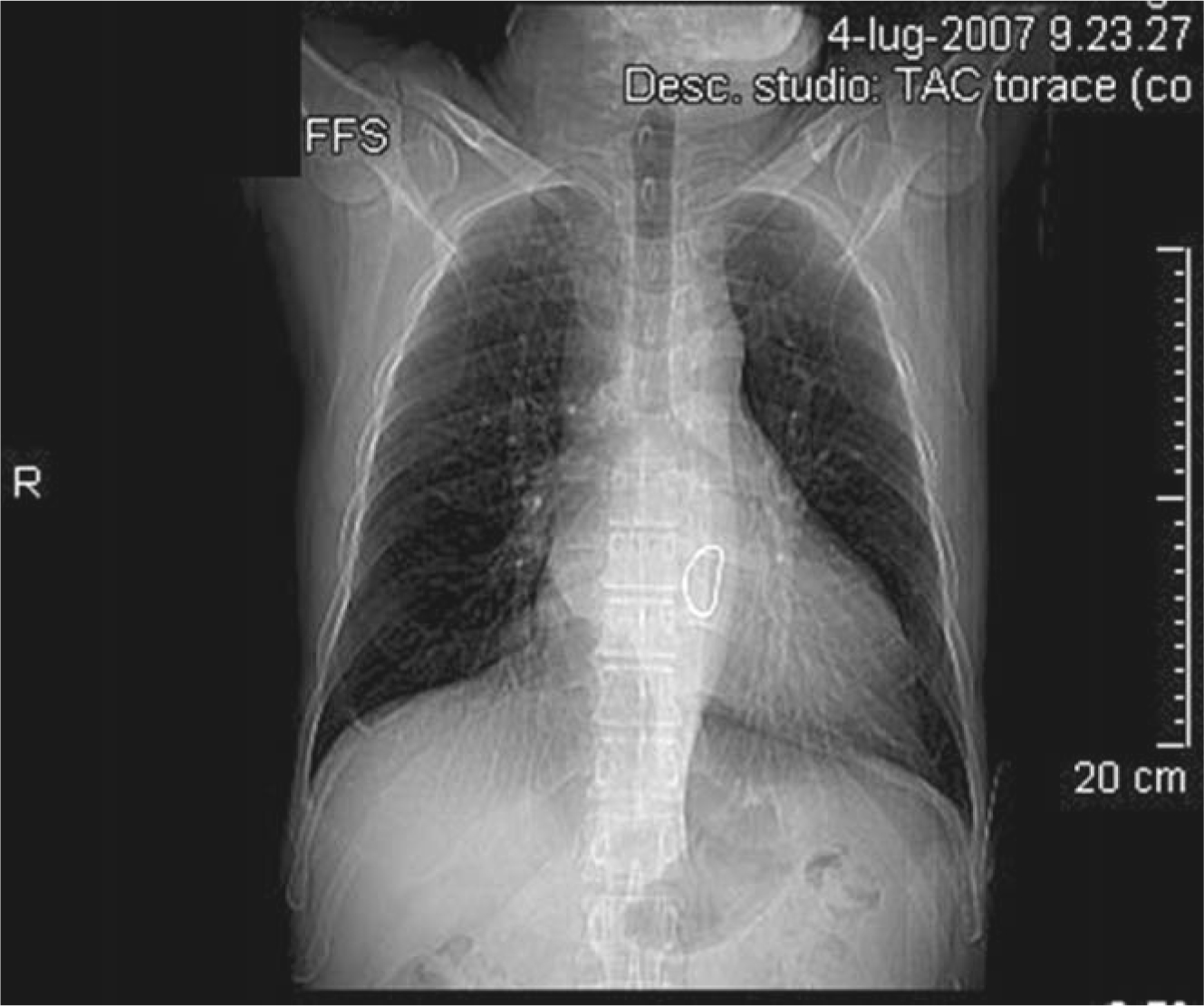 X Ray Anatomy Of The Heart Springerlink

 X Ray Anatomy
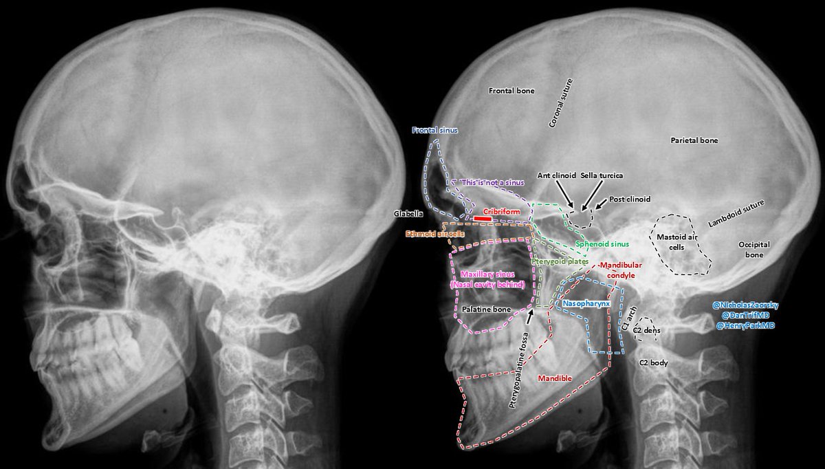 Nicholas Zaorsky Md Ms On Twitter Skull X Ray Anatomy For
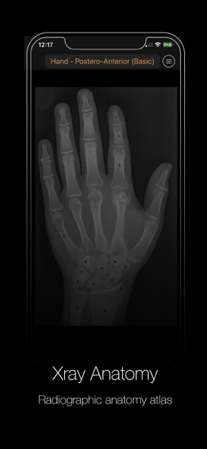 Xray Anatomy
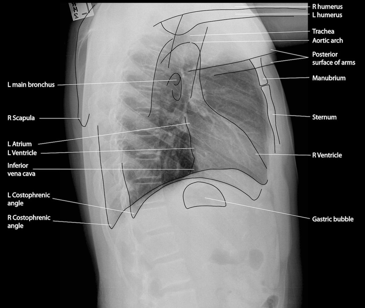 Normal Chest X Ray Litfl Medical Blog Labelled Radiology
![]() Medical Imaging And Radiological Anatomy X Ray Ct Mri
Medical Imaging And Radiological Anatomy X Ray Ct Mri
 Labeled Cervical Spine Xray Anatomy Lateral View Anatomy
 Normal Chest X Ray Terminology And Radiographic Anatomy
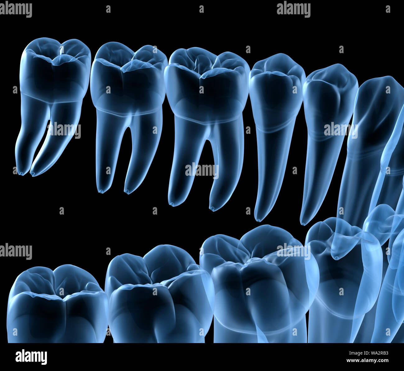 Dental Anatomy Of Mandibular Human Gum And Teeth X Ray View
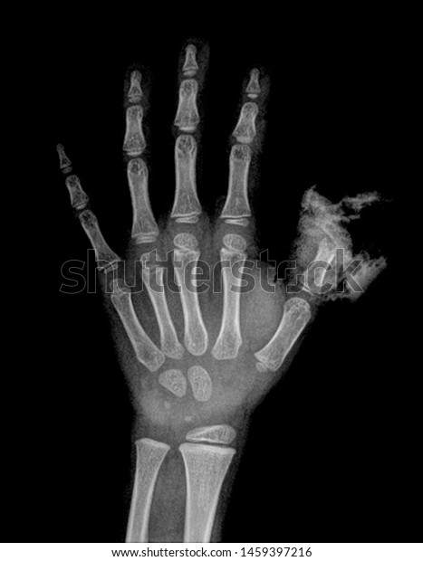
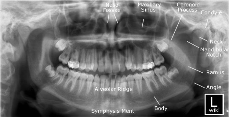 Mandible X Rays
Realistic Human Skeleton X Ray Anatomy Medical

 Radiographs Of The Dog
 Mandible X Rays
 Skeletal Anatomy 4 And An X Ray Image Of A Hand 5
 X Thorax Startradiology

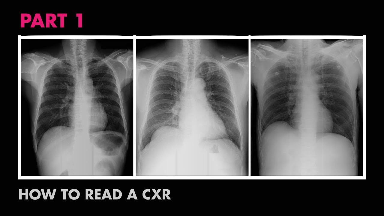 Anatomy Of A Chest X Ray How To Read A Chest X Ray Part 1
 Film Critique Part 1 Chest
 852 Calcaneus And Foot Anatomy The Xray Shows A Lateral
 Radiographic Anatomy Pelvis Ap Female My Next Practical I
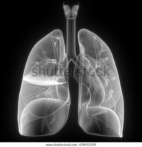 Human Body Organs Lungs Anatomy Xray Stock Illustration
 Lobar Cxr Anatomy
 Picture Show Rib Cage X Ray Image Of The Chest Showing

Belum ada Komentar untuk "Anatomy X Ray"
Posting Komentar