Ankle Anatomy Medial
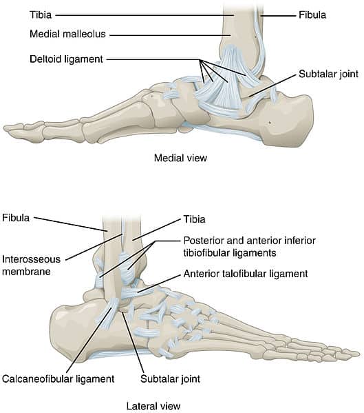
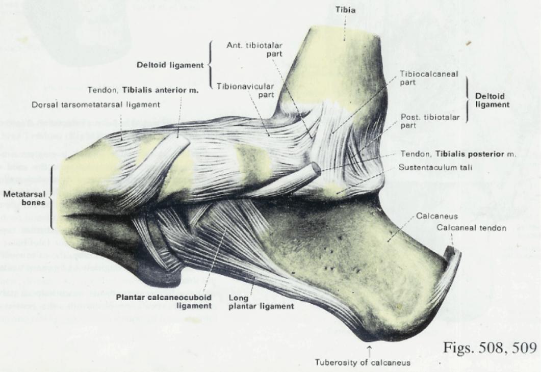
Medial ankle stability is provided by the strong deltoid ligament the anterior tibiofibular ligament and the bony mortise. The syndesmosis of the ankle refers to the membrane connecting the tibia to the fibula.
 The Fasciae Around The Ankle Human Anatomy
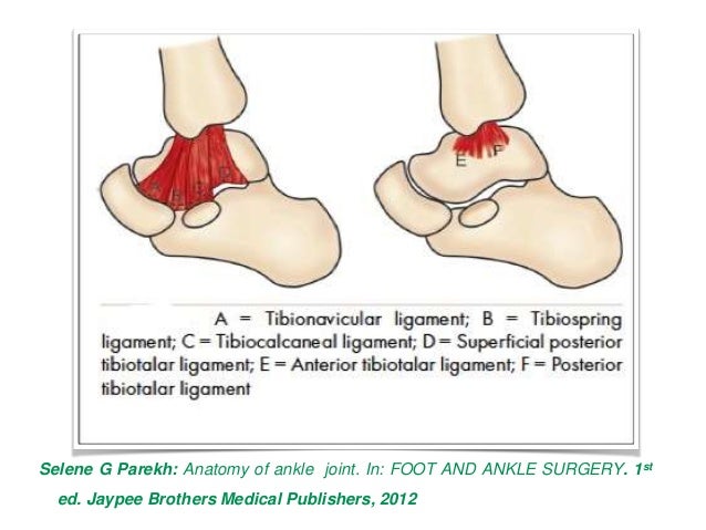
The medial collateral or deltoid ligament and lateral collateral ligament.

Ankle anatomy medial. The bones of the foot and ankle begin with the ankle joint itself. The calcaneofibular ligament cfl which connects the calcaneus or heel bone to. The lateral malleolus felt on the outside of your ankle is the low end of the fibula.
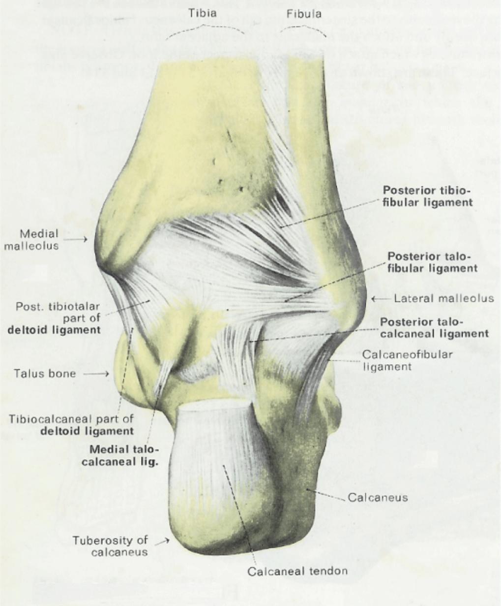
The lateral group of ankle ligaments composed of 3 ligaments. The ankle joint allows up and down movement of the foot. The lower ankle joint is formed by the talus calcaneus and navicular bone.
Lateral side of the ankle joint capsule. The upper ankle joint is formed by the inferior surfaces of tibia and fibula and the superior surface of talus. Three ligaments on the outside of the ankle that make up the lateral ligament complex as follows.
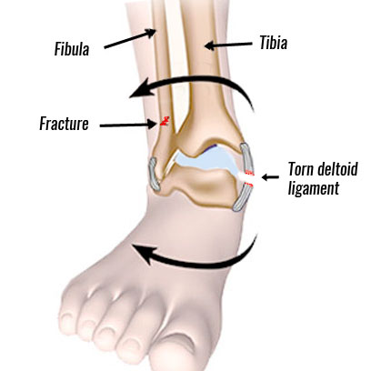
The anterior talofibular ligament atfl which connects the front of the talus bone to the fibula or shin bone. Because of the bony articulation between the medial malleolus and the talus medial ankle sprains are less common than lateral sprains. In medial ankle sprains.
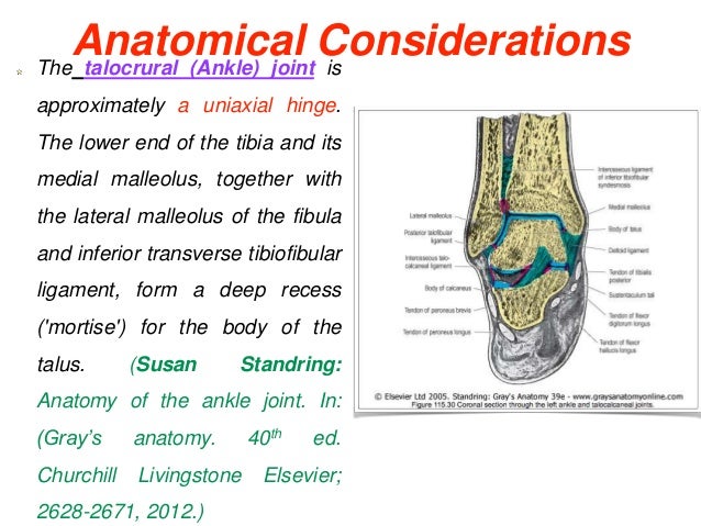
Calcaneofibular ligament anterior talofibular ligament and posterior talofibular ligament. The joint is supported by a set of ankle ligaments. Biomechanically a certain amount of motion is allowed in all planes with respect to the distal ends of the tibia and fibula.
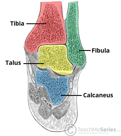
The ankle joint talocrural joint is formed where the distal end of the leg meets the foot. The ankle joint is formed where the talus the uppermost bone in the foot and the tibia shin meet. They attach to the lateral malleolus and they are smaller than the medial ligament which makes sprains of the lateral ligament to be more common.
The tibia and fibula are connected throughout their length by an interosseous membrane.

 Medial Ankle Pain
 Muscles And Tendons Of The Foot And Ankle Myfootshop Com
 The Ankle Joint Articulations Movements Teachmeanatomy
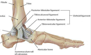
 Medial Ankle Ligament Physiopedia
 The Ankle Joint Articulations Movements Teachmeanatomy

 Ankle Sprain Medial Ligament Ankle Injury Physioadvisor
 Tendinopathies Of The Foot And Ankle American Family Physician
 Posterior Tibial Tendon Insufficiency Ptti Foot Ankle
Ankle Anatomy
 Eversion Ankle Sprain Medial Ankle Sprain

 Pin On Foot Issues
 Anatomy Of The Medial Ankle Foot
 Normal Anatomy Of The Medial Ankle Download Scientific
 Medial Ankle Pain
 11 Surface Anatomy Of The Medial Ankle Mm Medial Malleolus
 Ankle Anatomy
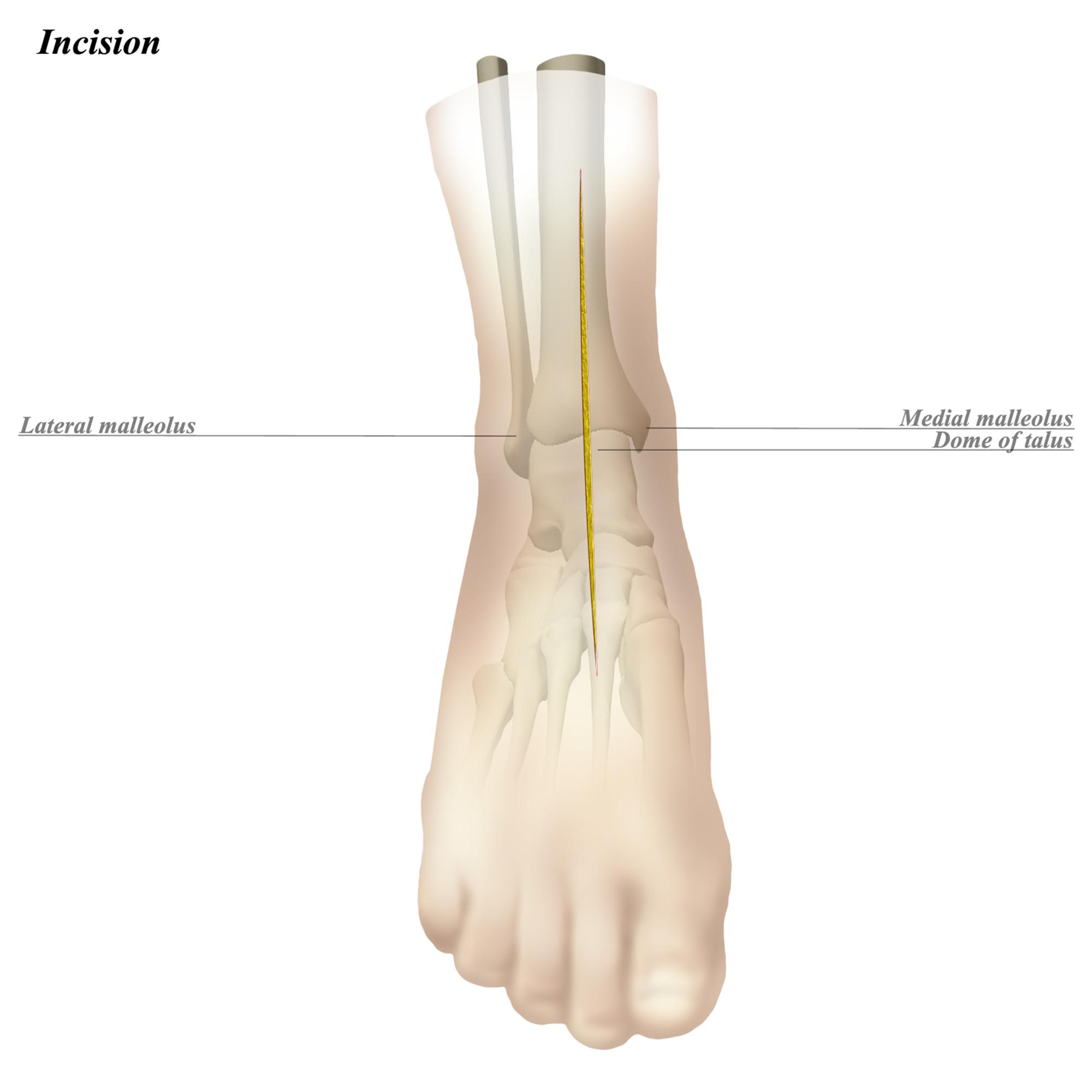 Ankle Anterior Approach Approaches Orthobullets
 Pin On Medical Info
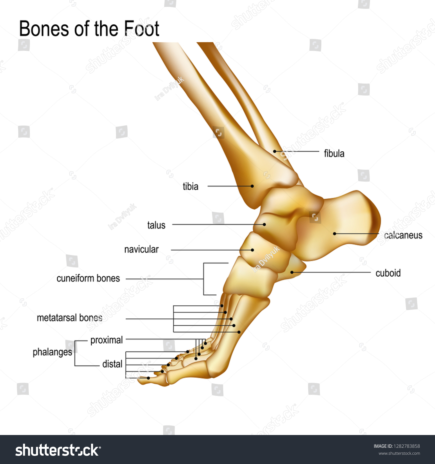 Foot Ankle Realistic Skeleton Human Leg Stock Vector
 Ankle And Foot Injuries Trauma Harwood Nuss Clinical
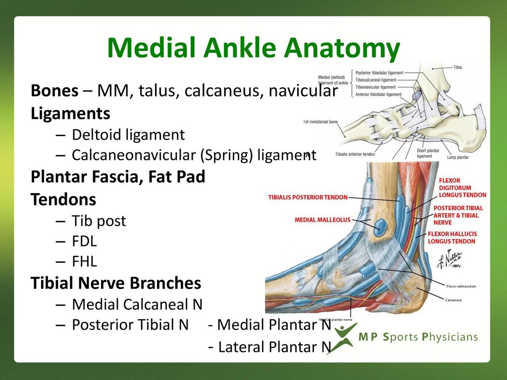 Medial Ankle And Heel Pain Ppt Download
 Pin On Doctors Anatomy Posters
 Duke Anatomy Lab 2 Pre Lab Exercise
 Medial Ankle Ligament Physiopedia
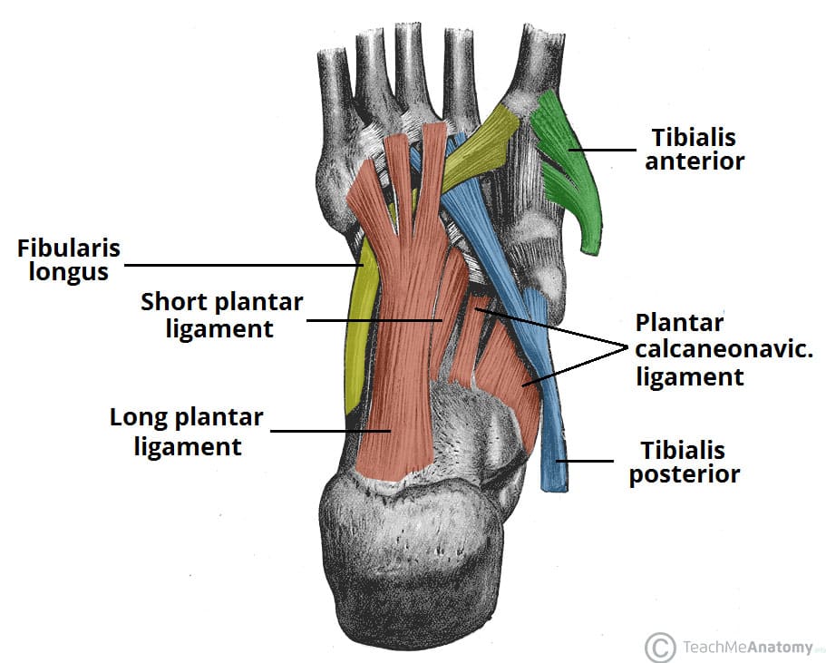 The Arches Of The Foot Longitudinal Transverse
Belum ada Komentar untuk "Ankle Anatomy Medial"
Posting Komentar