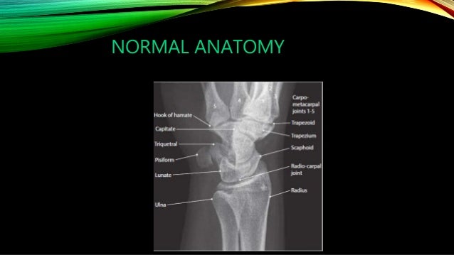
Hand Xray Anatomy
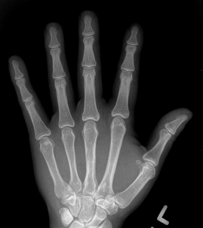
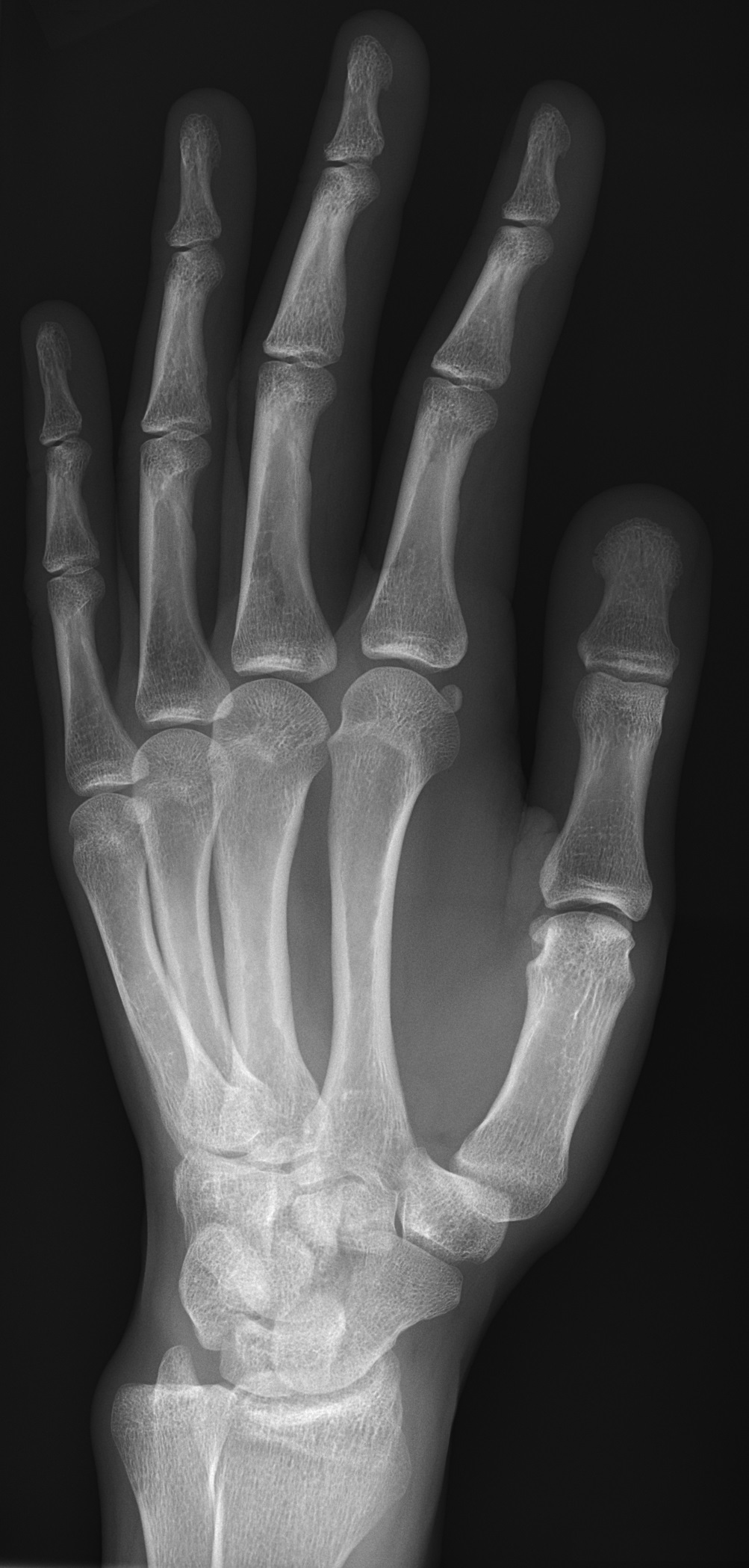
The hand comprises the metacarpal and phalangeal bones. Fractures and dislocations are usually straightforward to identify so long as the potentially injured bone is fully visible in 2 planes.
 X Hand Startradiology
For each image you will have four possible answers to choose from.

Hand xray anatomy. Adjustments in kvp and mas should be considered in cases involving splints casts wraps swelling braces etc. The quizzes below each have 15 multiple choice identification questions related to bones of the hand and foot and includes the following bones. Start studying hand xray anatomy.
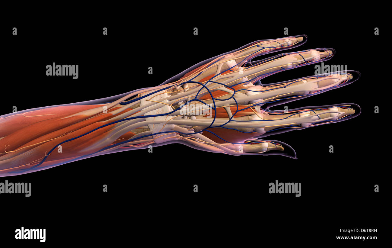
This is an interactive game to test your knowledge on the anatomy of the upper limb. How to interpret a chest x ray lesson 2 a systematic method and anatomy duration. A hand is a prehensile multi fingered appendage located at the end of the forearm or forelimb of primates such as humans chimpanzees monkeys and lemursa few other vertebrates such as the koala which has two opposable thumbs on each hand and fingerprints extremely similar to human fingerprints are often described as having hands instead of paws on their front limbs.
The x ray beam will pass through the hand from dorsal to palmar fig. Learn vocabulary terms and more with flashcards games and other study tools. For a pa image the hand lies flat on the x ray plate at the level of the shoulder with the elbow in 90 degrees flexion.
Quizzes on the bones of the hand and foot. The series primarily examines the radiocarpal and distal radioulnar joints the carpals metacarpals and phalanges. Hand anatomy animated tutorial duration.
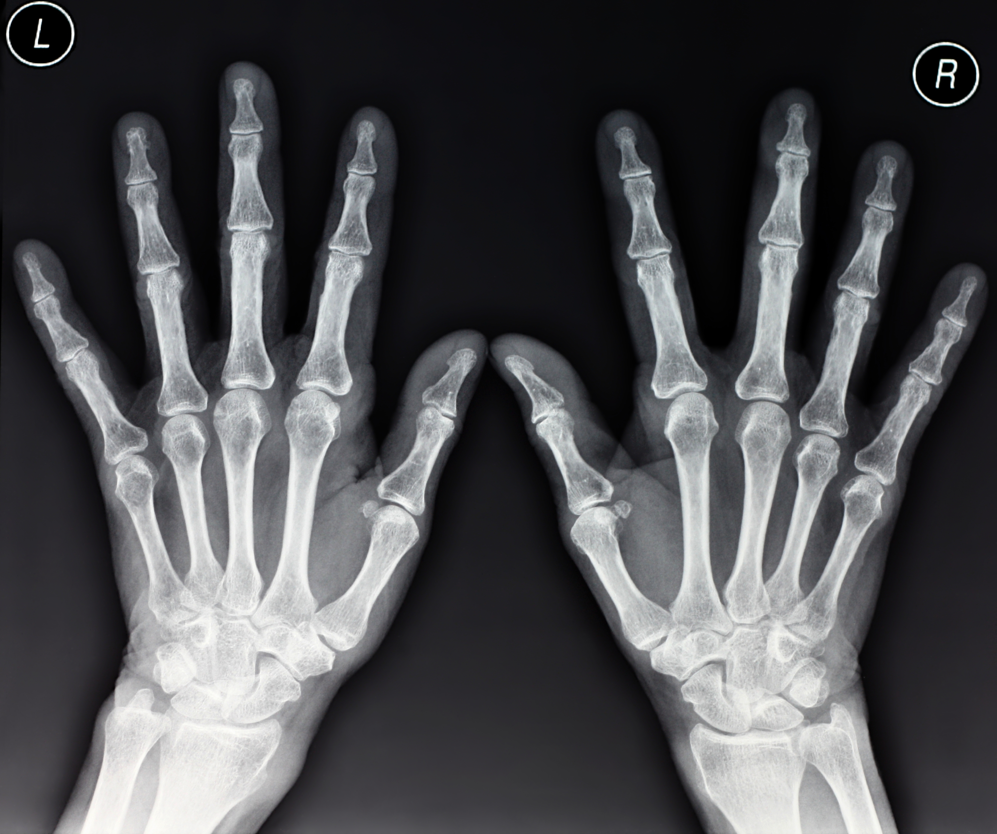
Finger injuries visible on x ray include bone fractures dislocations and avulsions. Articles here youll find a range of short articles on basic anatomy and physiology topics. Randale sechrest 659867 views.
Test your knowledge about hand xray anatomy with this online quiz. X ray games anatomy upper limb pictures quiz. Hand x rays are indicated for a variety of settings including.
A trivia quiz called hand xray anatomy. In this picture game you will label the structures of the upper limb from a series of x rays. Keep the body part as close to the cassette as possible in order to reduce oid object image distance.
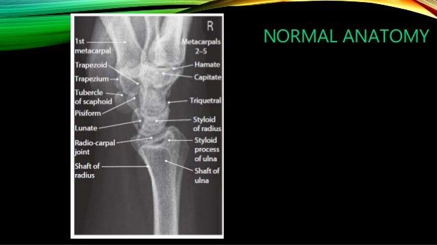
Although x ray machines vary the general kvp ranges for radiography of the wrist and hand is between 50 65 kvp. The hand series consists of a posteroanterior oblique and a lateral projectionalthough additional radiographs can be taken for specific indications. The standard examination of the hand generally consists of a posterior anterior pa image and a pa oblique image 34 image.
Trauma x ray upper limb handfingers.
 Female Hand And Wrist Anatomy Back Posterior View Xray
 Hand Radiographic Anatomy Wikiradiography
 Uams Gross Anatomy X Ray Atlas
 X Ray Of Wrist And Hand
Xray Image Of Normal Hand Xray Medical Background Stock
 Amazon Com Ahawoso Canvas Prints Wall Art 16x16 Inches
 Hand Xray Anatomy Purposegames
Baby S Hand X Ray Stock Image P116 0845 Science Photo
 Radiology Anatomy
Radiology Of The Hand
 X Hand Startradiology
 Xray Right Hand Finger Image Photo Free Trial Bigstock
 Abc Of Emergency Radiology The Wrist The Bmj
 Basilar Thumb Arthritis Hand Orthobullets
 Radiographic Anatomy Of Adult Hand Orthopaedicsone
 Hand Finger Thumb Hospital Xray Scan Stock Image Image Of
 Image Left Hand X Ray X Ray Image Of Male Human Left Hand
 What Does Hand Arthritis Look Like On X Rays Raleigh Hand
Stock Illustration Human Wrist Anatomy Xray View
 The Radiology Assistant Wrist Carpal Instability
 X Ray Of Wrist And Hand
 Radiographic Anatomy Wrist Ap Medical Radiography
 Skeletal Trauma
 The Importance Of Radiopaque Markers In Digital X Ray
 X Hand Startradiology
 File X Ray Of Normal Hand By Oblique Projection Jpg
 Hand Bone Anatomy X Vector Photo Free Trial Bigstock


Belum ada Komentar untuk "Hand Xray Anatomy"
Posting Komentar