Knee Anatomy Xray
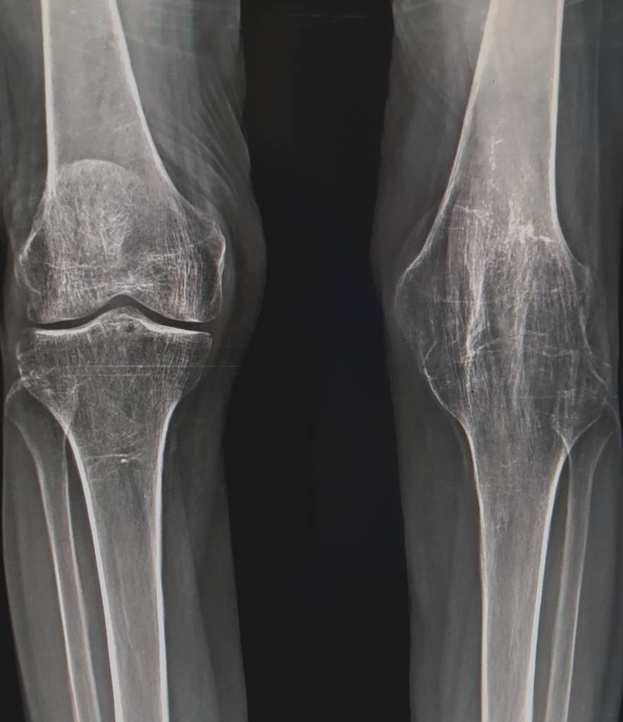
Click on a link to get t1 coronal view t2 fatsat axial view t2 fatsat coronal view t2 fatsat sagittal view. This is a front to back view of the knee joint also called the ap view.
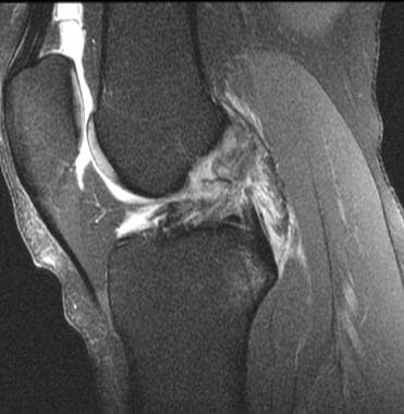
 Ankylosis Of The Knee Joint Radiology Case Radiopaedia Org
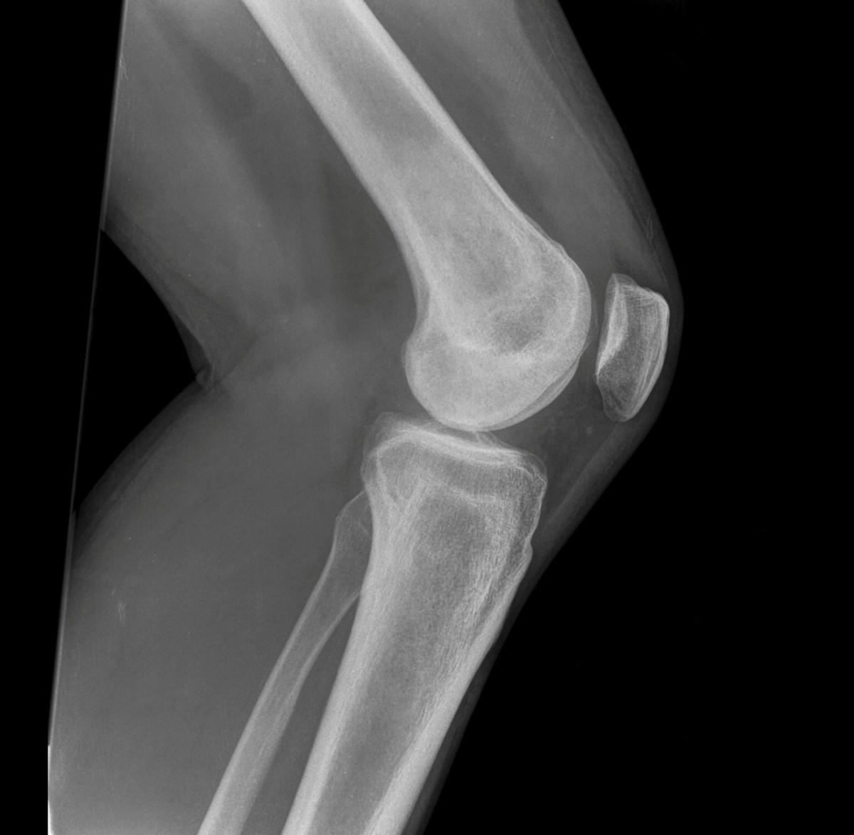
A standard examination includes an anterior posterior image and a lateral image.

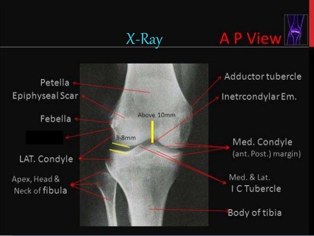
Knee anatomy xray. Stanford bone tumor bayesian network issssr msk lectures for residents ocad msk cases from around the world stanford msk mri atlas has served almost 800000 pages to users in over 100 countries. Use the mouse to scroll. It is the largest synovial joint in the body and allows flexion and extension of the leg as well as some rotation in the flexed position.
Similar to the medial aspect of the knee the lateral knee supporting structures are described in layers from superficial to deep figure 13 2. A fracture here is most common in adolescents following hyperextension of the knee. This allows effusions to be visualised in the suprapatellar pouch.
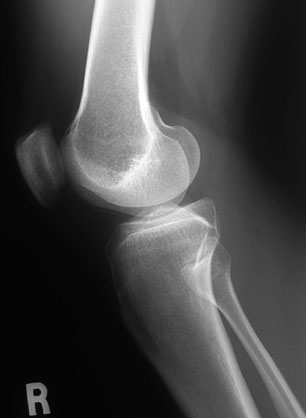
An x ray is one of the most common imaging tests used to diagnose a knee problem. Anterior cruciate ligament acl is one of the two cruciate ligaments that stabilize the knee joint. In the context of trauma the lateral view is acquired with the patient lying supine and with a horizontal x ray beam.
Knee normal ap. The iliotibial band is a vertically oriented ligamentous fascia that attaches to gerdy tubercle that is located at the anterolateral aspect of the proximal tibia. Its an avulsion fracture at the tibial attachment of the acl.
This webpage presents the anatomical structures found on knee mri. Ap stands for anteroposterior meaning the image is directed from the front to the back of the knee joint. The knee joint is a modified hinge joint between the femur tibia and patella.
To establish the presence of a fracture as in each conventional x ray the knee should be imaged in at least two directions. Gross anatomy the acl arises from the anteromedial aspect of the intercondylar area on the tibial plateau and passes upwards and backwards to. Atlas of knee mri anatomy.
 Mri For Anterior Cruciate Ligament Injury Overview Anatomy
 Lower Limb Radiographs
 Vector Illustration Anatomy Front X Ray Of A Healthy Knee Joint
 Radiographic Anatomy Of The Skeleton Knee Lateral View
 Knee Xray Bones Human Leg Anatomy Stock Photo Download
 X Ray Bones The Of Knee Stock Vector C Iradvilyuk 274337944
Paediatric Knee Wikiradiography
 Mri Knee Joint Anatomy
Knee Non Trauma Radiographic Anatomy Wikiradiography
 Chapter 7 Imaging Of Joints Basic Radiology 2e
 Common Mistakes And Pitfalls In Magnetic Resonance Imaging
 Test Yourself Regular Sets Set 27 Emergency Department
Anatomy Hip Knee Book
 Radiological Anatomy Of The Lower Limb
 Amazon Com Canvas On Demand Arthritis Of The Knee X Ray
 Vector Illustration Anatomy Of A Healthy Knee Joint Front X Ray
 Knee Series Radiology Reference Article Radiopaedia Org
 Knee Anatomy The Knee Is The Largest Joint In The Body The
 Vector Illustration Anatomy Of A Healthy Knee Joint Front
 Xray Knee Joint Anatomy Diagram Quizlet
 Human Knee Anatomy Lateral View Canvas Print
 Ecr 2017 C 0585 All About The Patella Not Just A Cap
 Anatomy Of The Knee Ct Arthrography
Xray Knee Stock Photo More Pictures Of Anatomy Istock
 Anatomy Of The Knee Joint Owlcation
 Knee Leg Atlas Of Anatomy
 Normal Radiographic Anatomy Of The Knee Radiology Case
 Knee Anatomy Xray View Medically Accurate 3d Illustration
 Film Knee X Ray Radiograph Show Normal Human Anatomy Of Knee
 Lower Limb Radiographs
 Human Knee Anatomy In X Ray Buy This Stock Illustration
 Full Text Radiofrequency Techniques To Treat Chronic Knee
 Arteries And Bones Of The Lower Extremity Interactive Atlas
 Radiography Knee Injury And Prevention


Belum ada Komentar untuk "Knee Anatomy Xray"
Posting Komentar