Anatomy Of The Skull And Neck
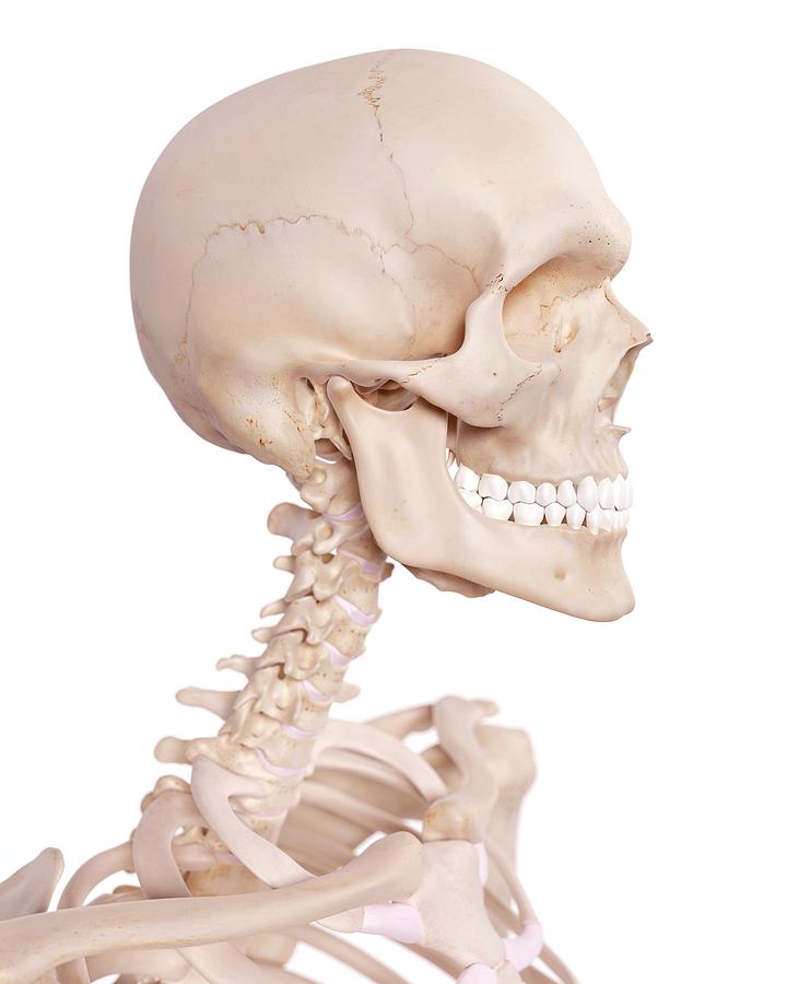
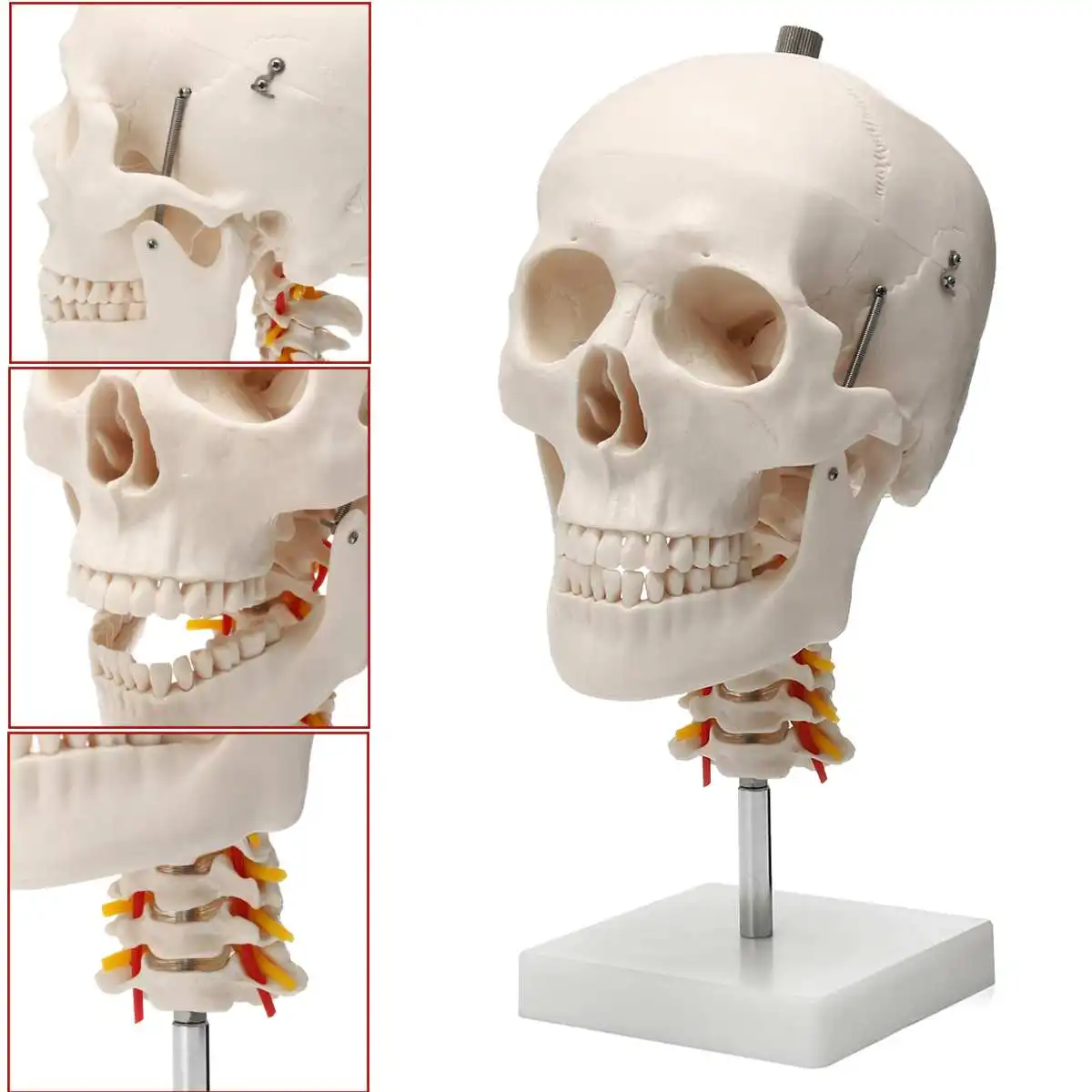
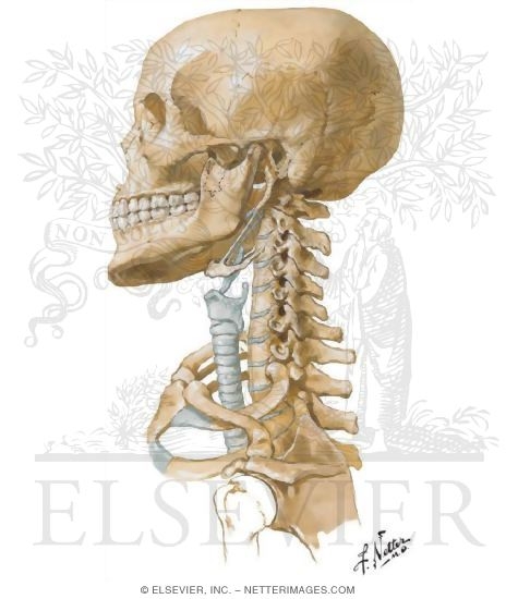
The head rests on the top part of the vertebral column with the skull joining at c1 the first cervical vertebra known as the atlas. The human head weighs nearly as much as the average bowling ball at around ten to twelve pounds respectively.
 General Anatomy Of The Bull And The Cow Illustrated Atlas
Despite being a relatively small region it contains a range of important anatomical features.

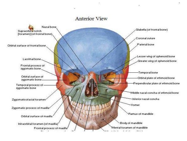
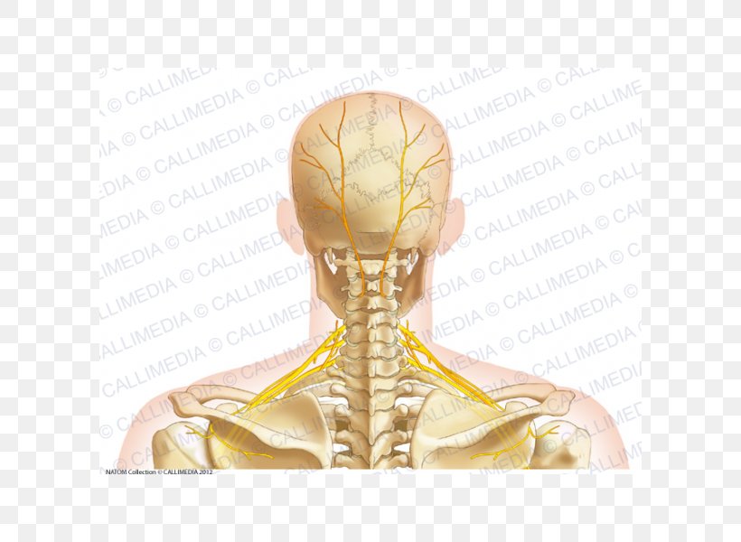
Anatomy of the skull and neck. The occipital bone is the only bone in your head that connects with your cervical spine neck. Anatomy of the skull the skull figures 21 and 22 has two defined areas. One of the functions of the neck is to act as a conduit for nerves and vessels between the head and the trunk.
The foramen magnum allows key nerves and vascular structures passage between the brain and spine. If you continue to use this site we will assume that you are happy with it. The cranial and facial bones and structures.
There are eight bones that make up the cranium. Think of it like a jigsaw puzzle all the pieces fit in together and are required to get the full picture as to how it works. The occipital bone surrounds a large opening known as the foramen magnum.
It is made up of bones discs muscles ligaments nerves and tendons. Detailed anatomy of the human skull. Radiological anatomy of the head and neck on a ct in axial coronal and sagittal sections and on a 3d images.
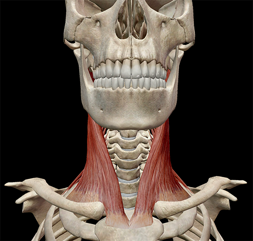
They move the head in every direction pulling the skull and jaw towards the shoulders spine and scapula. Namely it is what the spinal cord passes through to enter the skull. The cranium and facial.
The single bones are the frontal occipital sphenoid and ethmoid and the paired bones are the parietal and temporal. From supporting the head to containing the spinal cord and nerves as they emerge from the skull this structure does it all. We use cookies to ensure that we give you the best experience on our website.
The skeletal section of the head and neck forms the top part of the axial skeleton and is made up of the skull hyoid bone auditory ossicles and cervical spine. The neck muscles including the sternocleidomastoid and the trapezius are responsible for the gross motor movement in the muscular system of the head and neck. Neck anatomy pictures bones muscles nerves.
The neck begins at the base of the skull and connects to the thoracic spine the upper back. The neck has the ability to support a great deal of weight too. Anatomy of the head and neck ct scan ct scan of head and neck.
The neck is the area between the skull base and the clavicles.
 Human Skull And Cervical Spine
 Free Anatomy Quiz The Human Skull Quiz 1
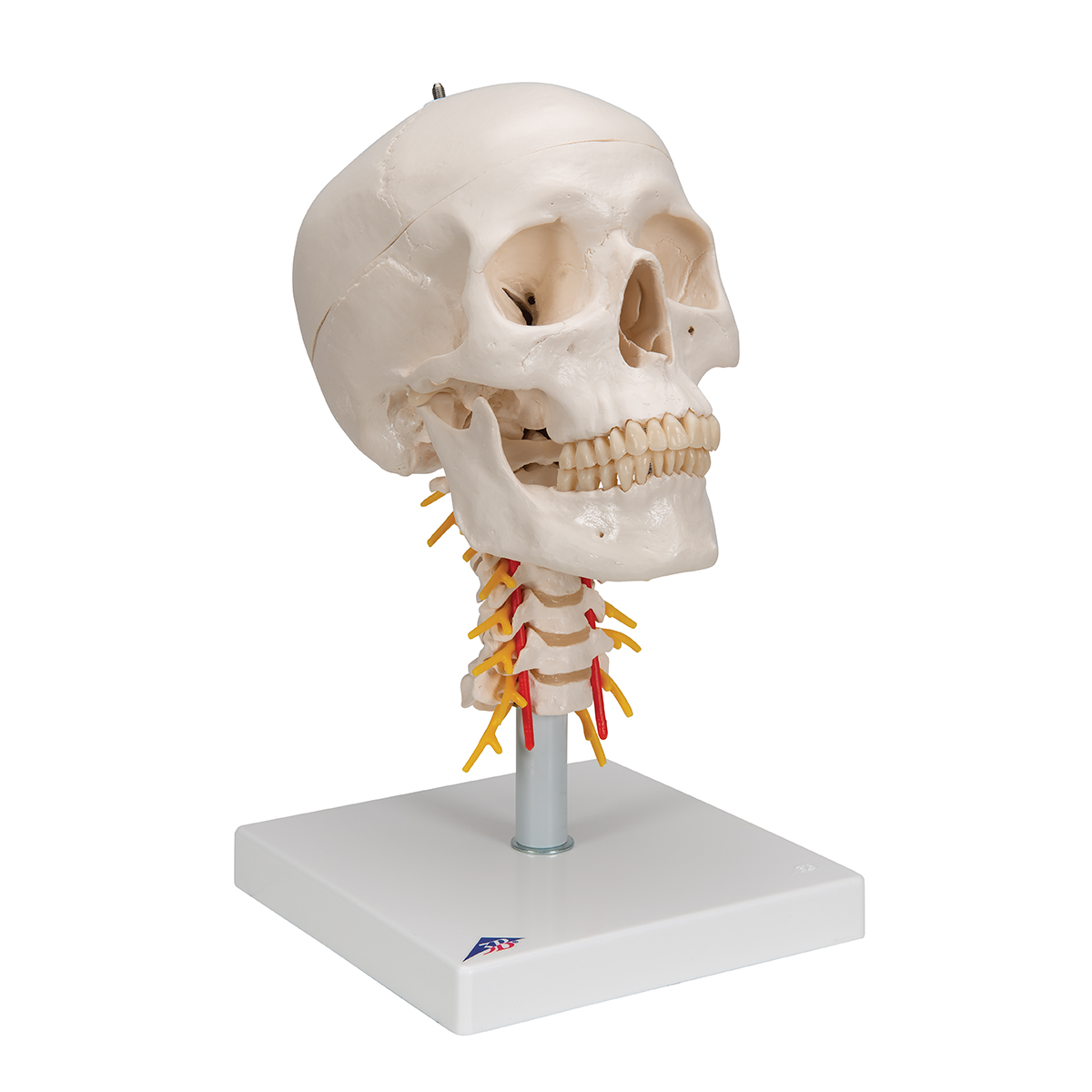
 Classic Human Skull Anatomy Model On Cervical Spine
 Occipital Neuralgia
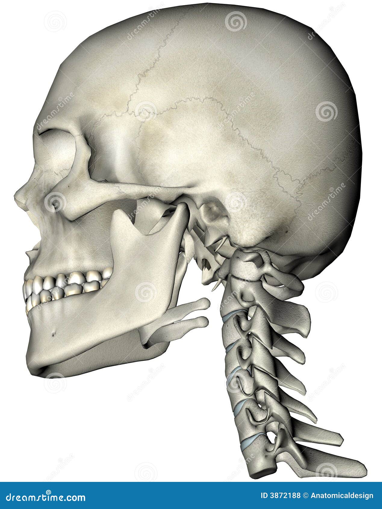 Human Skull And Neck Lateral Stock Illustration
The Skull Anatomy And Physiology Openstax
 Learn Muscle Anatomy Scalene Muscles And Other Neck Anatomy
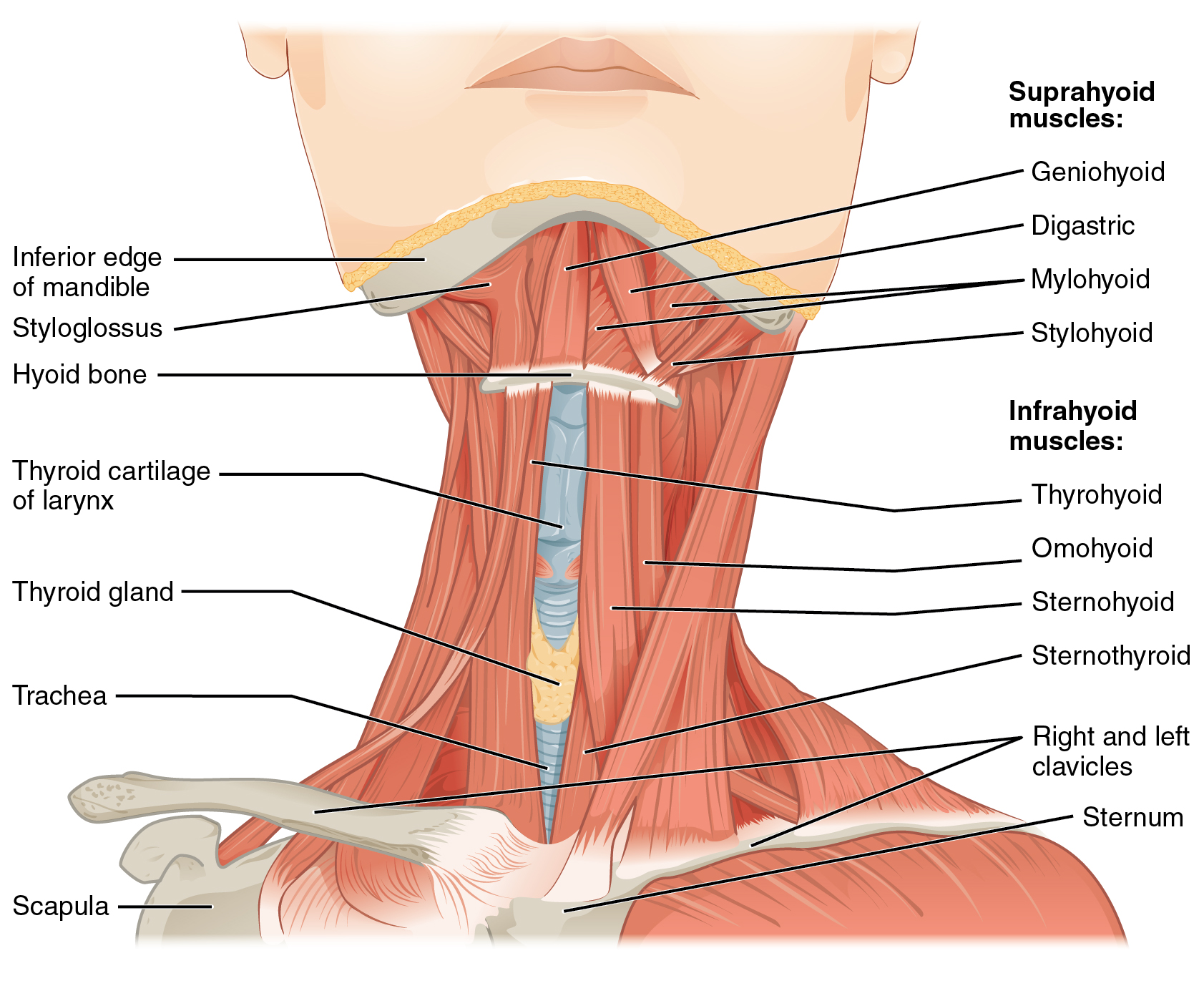 11 3 Axial Muscles Of The Head Neck And Back Anatomy And
 Neck Cancer Anatomy Headandneckcancerguide Org

 Head And Neck Bone Nerves Anterior Image
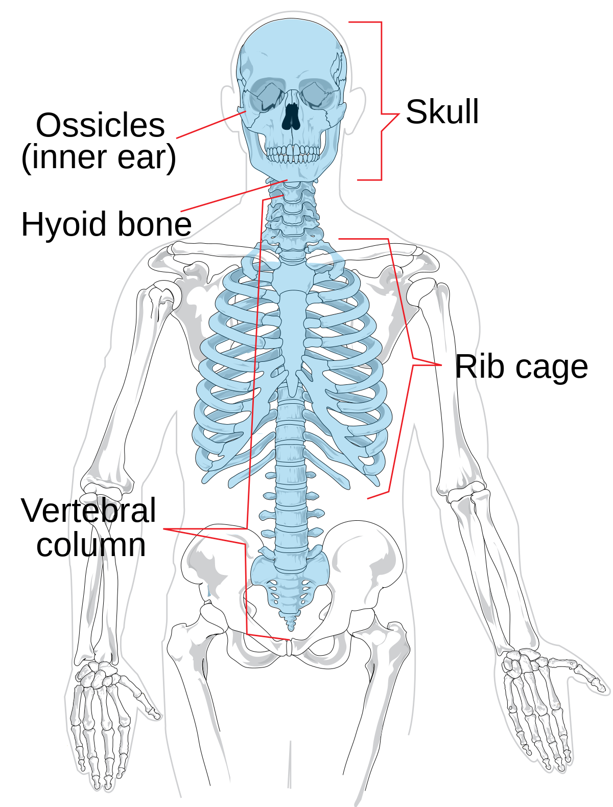 Axial Skeleton Wikipedia
 Neuroanatomy Head Model
 Gross Anatomy Of The Head And Neck
 Us 43 28 40 Off 1 1 Life Size Human Skull Anatomical Anatomy Skull Model Cervical Spine Head Skeleton School Educational Medical Teaching Model In
 Head Skull And Neck Anatomy
 Neck Bone Human Anatomy Head Png 600x600px Watercolor
 Massage Therapy For Tension Headaches
Anatomy Of Salivary Gland Cancer Headandneckcancerguide Org
 Head And Neck Anatomy

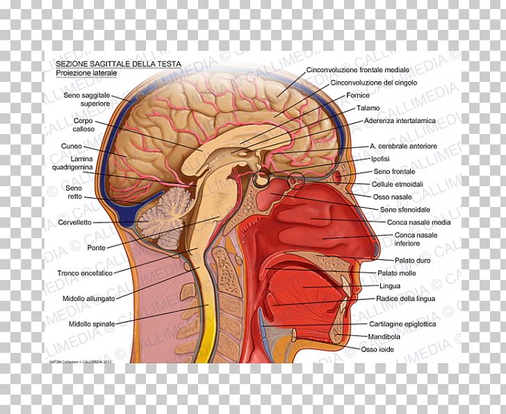 Sagittal Plane Human Head Brain Skull Png Clipart Anatomy
 Head And Neck Anatomy Head And Neck Anatomy Bone Human
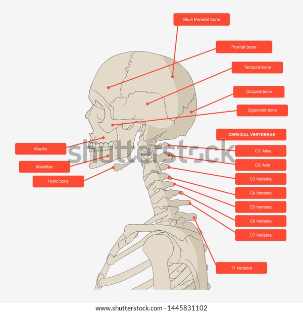 Lateral View Skull Neck Vertebrae Bones Stock Vector
 The Veins Of The Head And Neck Human Anatomy
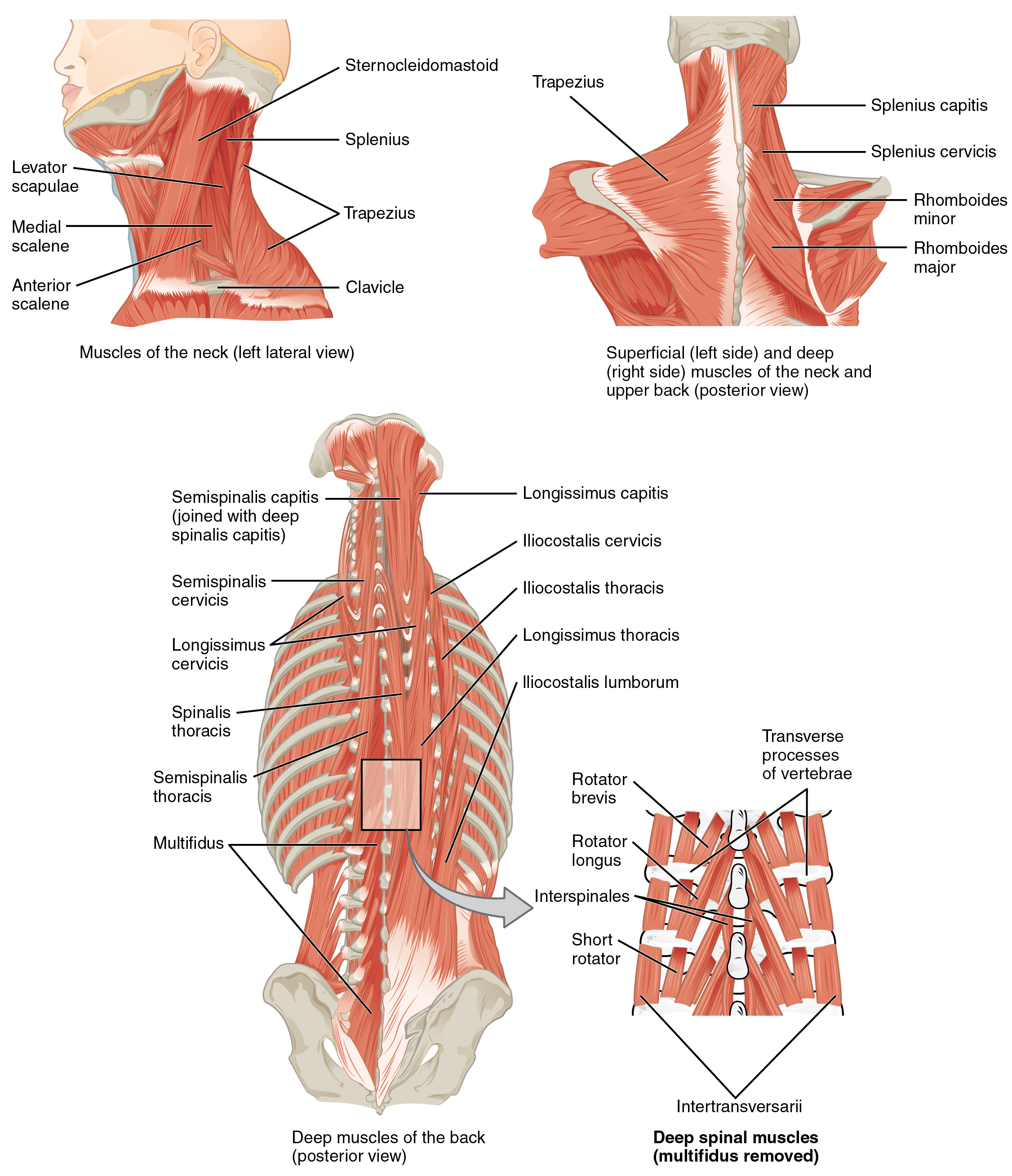 11 3 Axial Muscles Of The Head Neck And Back Anatomy And
 Human Skull Model Plastic Skull Model Human Skull Model
 Skull And Neck
 Cardiovascular System Of The Head And Neck
 Nerves Of The Head And Neck Interactive Anatomy Guide
Belum ada Komentar untuk "Anatomy Of The Skull And Neck"
Posting Komentar