Ear Canal Anatomy
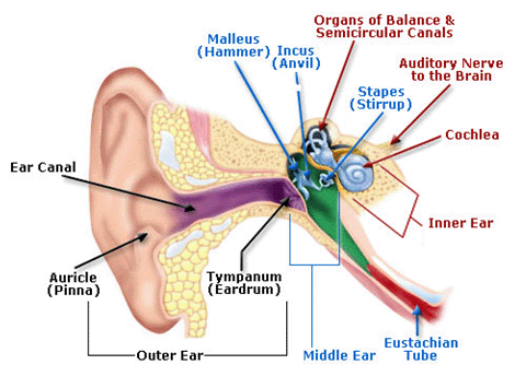
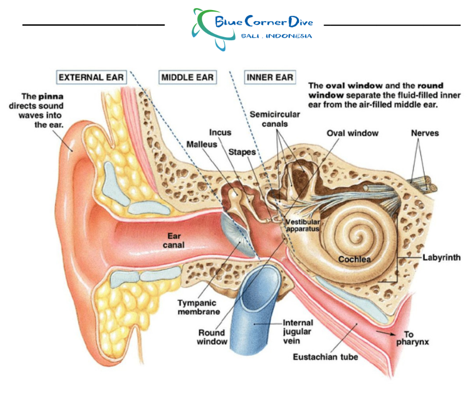
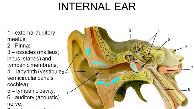
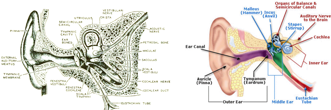
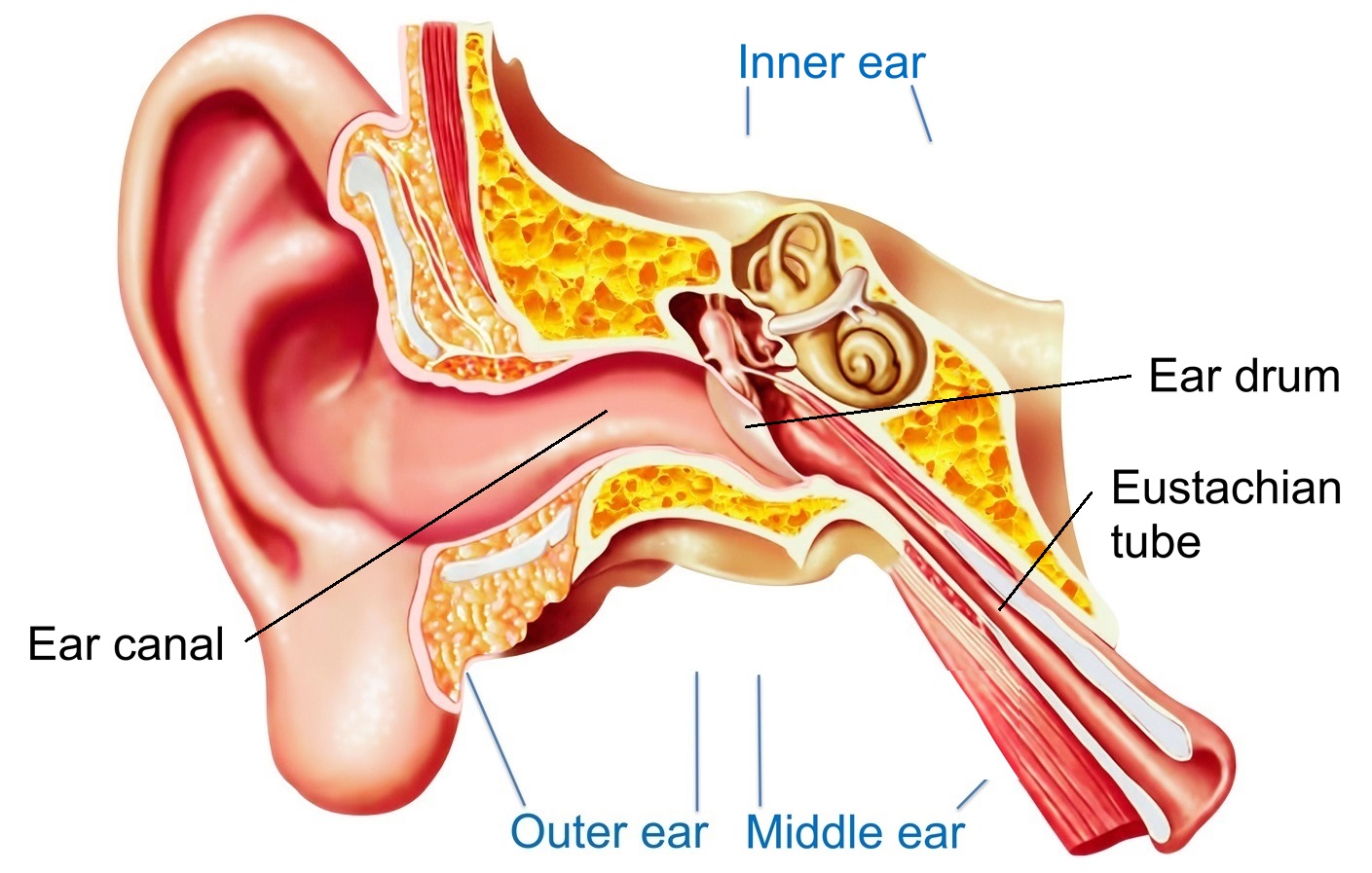
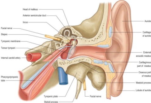
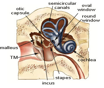
The middle ear contains three small bones ossicles that transmit sound waves from the eardrum to the inner ear. The ear canal also called the external acoustic meatus is a passage comprised of bone and skin leading to the eardrum.
The skin of the ear canal is very sensitive to pain and pressure.

Ear canal anatomy. Ear canal the ear canal starts at the outer ear and ends at the ear drum. It travels from the outer ear to the eardrum. The ear canal external acoustic meatus external auditory meatus eam is a pathway running from the outer ear to the middle ear.
The ear canal contains protective hairs and ear wax cerumen. The canal is approximately an inch in length. Sound travels through the auricle and the auditory canal a short tube that ends at the eardrum.
External auditory canal or tube. Auricle cartilage covered by skin placed on opposite sides of the head auditory canal also called the ear canal eardrum outer layer also called the tympanic membrane the outer part of the ear collects sound. The translucency of the tympanic membrane allows the structures within the middle ear to be observed during otoscopy.
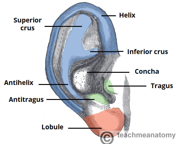
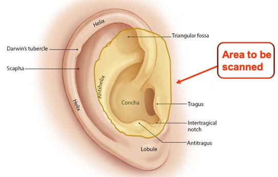
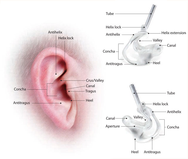
Tympanic membrane also called the eardrum. The outer ear is called the pinna and is made of ridged cartilage covered by skin. External or outer ear consisting of.
This is the outside part of the ear. The average length of the canal is 26 mm 102 inches with a diameter of 7 mm about 025 inches. The three bones are called the malleus the incus and the stapes.
The ears canal is a small tube located in the middle ear that connects the outer ear to the inner ear. The ear is comprised of the ear canal also known as the outer ear the. The middle ear tympanic cavity lies behind the eardrum.
The actual size varies with each individual. The ear is the organ of hearing and balance. This is the tube that connects the outer ear to the inside or middle ear.
The outer ear includes. Bony lesions of ear canal. The parts of the ear include.
These bony lesions can generally be managed with vigilant cleaning of ear wax to prevent obstruction. On the inner surface of the membrane the handle of malleus attaches to the tympanic membrane at a point called the umbo of tympanic membrane. Ear anatomy outer ear.
Sound funnels through the pinna into the external auditory canal a short tube that ends at the eardrum tympanic. These are benign growths of bone along the walls of the ear canal resulting in a narrowing of the ear canal which may then lead to frequent obstruction from a small amount of wax or water. Under the skin the outer one third of the canal is cartilage and inner two thirds is bone.
The adult human ear canal extends from the pinna to the eardrum and is about 25 centimetres 1 in in length and 07 centimetres 03 in in diameter.
 Ear Injuries Diving Everything You Need To Know Manta Dive
 Ear Structure And Function In Horses Horse Owners Merck
 Human Ear Canal Anatomy Detailed Illustration Wall Clock By Azza1070
 What Happens To My Ears When I Scuba Dive Blue Corner
 Tympanic Membrane Rupture And Middle Ear Infection In Cats
 The External Ear Structure Function Innervation
 Human Ear Structure And Anatomy Online Biology Notes
 The Moovin Groovin Outer Ear Lydia Gregoret Hearing Aids
 Ear Infection Middle Ear Symptoms Treatment Southern
 Otoscan 3d Ear Scanning The Future Is Now Jackie
 Swimmer S Ear Otitis Externa Is Inflammation Of The Ear Canal
 The Association Between Tinnitus The Neck And Tmj
 Blows To Head Can Damage Your Ears

Anatomy Of The Ear Diagnosis 101
A Beginner S Guide To Ear Anatomy And Physiology
Ear Anatomy Diagram Enchantedlearning Com
 Health Tips Archives Southern California Ear Nose Throat
 Pinna And External Auditory Canal Anatomy Springerlink


Belum ada Komentar untuk "Ear Canal Anatomy"
Posting Komentar