Sinus Cavity Anatomy
This lining secretes mucus a complex substance that keeps the nose and sinuses moist. Skip navigation sign in.

Or a cavity within a bone.

Sinus cavity anatomy. When they arent moistening the air we breathe through our noses. Clinical anatomy nasal cavity and sinuses duration. Treatment depends on the cause of the infection 40.
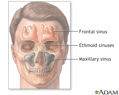
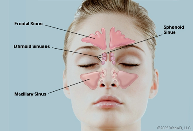
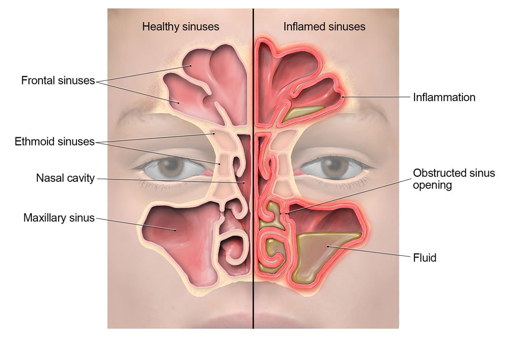
The frontal sinuses are in the lower center of the forehead bone above the eyes and nasal bridge. A viral bacterial or fungal condition it is characterized by a swollen and inflamed nasal cavity. Projecting out of the lateral walls of the nasal cavity are curved shelves of bone.
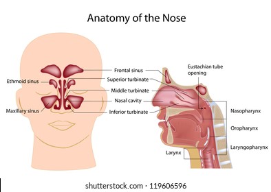
The sphenoid sinuses are behind the nasal. Frontal sinus cavities which can be found above the eyes more in the forehead region. The four paired sinuses or air cavities can be referred to as.
Armando hasudungan 297486 views. Mucus also helps trap dust viruses and bacteria and removes them from the nose. Also known as a sinus headache or sinusitis it causes pain and pressure in and around the sinuses forehead eyes and teeth 39.
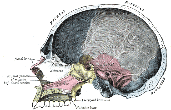
It is separated down the middle by the nasal septum a piece of cartilage which shapes and separates the nostrils. The low center of your forehead is where your frontal sinuses are located. Two types of sinus the blood filled and the air filled sinuses are discussed in this article.
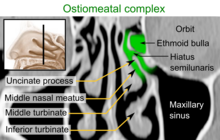
Each nostril can be further divided into roof floor and walls. They look like a mesh formation. The nasal cavity is the most superior part of the respiratory tract.
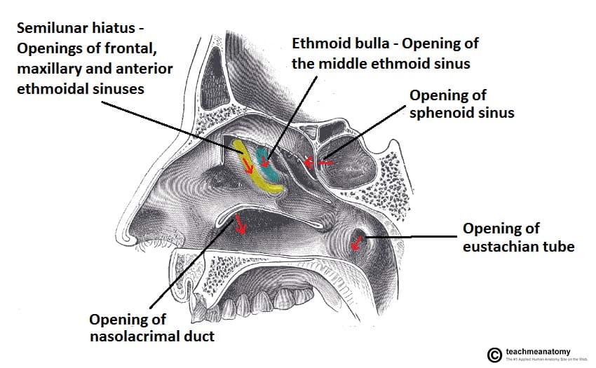
Openings into the nasal cavity. The sinuses are named for the bones where theyre located. Maxillary sinus cavities are located on either side of the nostrils cheekbone areas.
Nasal sinuses are covered with a special lining similar to the lining in the nasal cavity called mucosa. Sinus in anatomy a hollow cavity recess or pocket. Others are much smaller.
The nasal cavity divisions. Anatomy of the nasal cavitymov. The largest sinus cavities are about an inch across.
Anatomy of the nasal cavitymov. Your cheekbones hold your maxillary sinuses the largest. Between your eyes are your ethmoid sinuses.
Anatomy of the nasal cavity. The sinuses are a connected system of hollow cavities in the skull. The ethmoid sinuses are at the nasal bridge between the eyes.
The mucus secretions produced in the sinuses are continually being swept into the nose by the hair like structures called cilia on the surface of the respiratory membrane. One of the functions of the nose is to drain a variety. Ethmoid sinus cavities which are located between the eyes.
Like the nasal cavity the sinuses are all lined with mucus. The nasal cavity can be divided into the vestibule respiratory and olfactory sections. A large channel containing blood.
 Paranasal Sinuses An Overview Sciencedirect Topics
 Sinuses Picture Image On Medicinenet Com
 Understand Breathing And Sinus Expert Sinus Surgeon Nyc
 Equine Sinuses Ibook Demo
 The Nasal Cavity Structure Vasculature Innervation
Anatomy Of The Nasal Cavity
 Sinus Infection Sinusitis Symptoms Signs Treatment
 Sinus Cavities Paranasal Sinuses Location Anatomy
 The Nasal Cavity Structure Vasculature Innervation
 Pin By Sue Sparks On For Healthy Person Sinus Cavities
 Anatomy And The Human Blockhead Anatomy Of The Nasal
 Gross Anatomy Of Nasal Cavity And Paranasal Sinuses
 Science Source Nasal Cavity Illustration
 What Are The Sinuses Pictures Of Nasal Cavities

 Sinus Cavity Diagram Reading Industrial Wiring Diagrams

 Paranasal Sinuses An Overview Sciencedirect Topics
 Nasal Sinus Cavity Anatomy Sinus Cavities Paranasal
 Paranasal Sinuses Sinus Remedies Home Remedies For Sinus
 Nose And Sinus Cancer Anatomy Headandneckcancerguide Org
 Amazon Com Emvency Wall Tapestry Sinuses Of Nose Human
 Nose And Paranasal Sinuses Anatomy E Lab
 Nasal Cavity Images Stock Photos Vectors Shutterstock
 Nasal Cavity
 Nasal Cavity Wikipedia
 Patient Resource Publishing Head And Neck Sinus Nasal
 Anatomy Notes Balloon Sinuplasty
 Sinus Infection Stock Image C022 1583 Science Photo
Belum ada Komentar untuk "Sinus Cavity Anatomy"
Posting Komentar