Ct Anatomy Of The Head
Jakab m kikinis r. Adequate gray matter white matter differentiation.
 Ct Scan Of Head And Neck
Anatomy of the head and neck ct scan ct scan of head and neck.

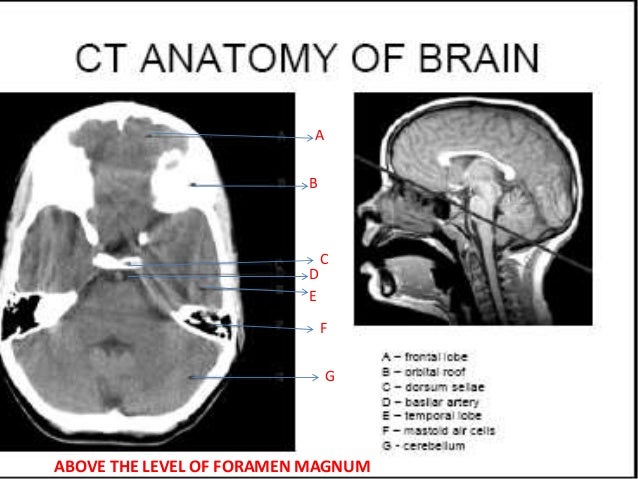
Ct anatomy of the head. The anterior part of the head is at the top of the image. This means that the right side of the brain is on the left side of the viewer. Spl head and neck atlas 2012 november.
The lambda is the point where joins lambdoid sutures and the sagittal suture. The coronal suture is the line where the parietal bone frontal bone and are in contact. The lambdoid suture is a line where the parietal bone occipital bone and are in contact.
The sagittal suture is the line where the right and left parietal bone are in contact. Ct head neck atlas. Welcome to online mri ct sectional anatomy.
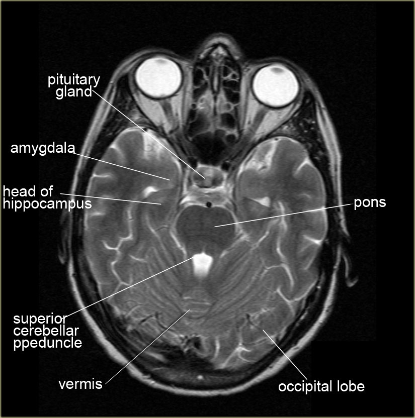
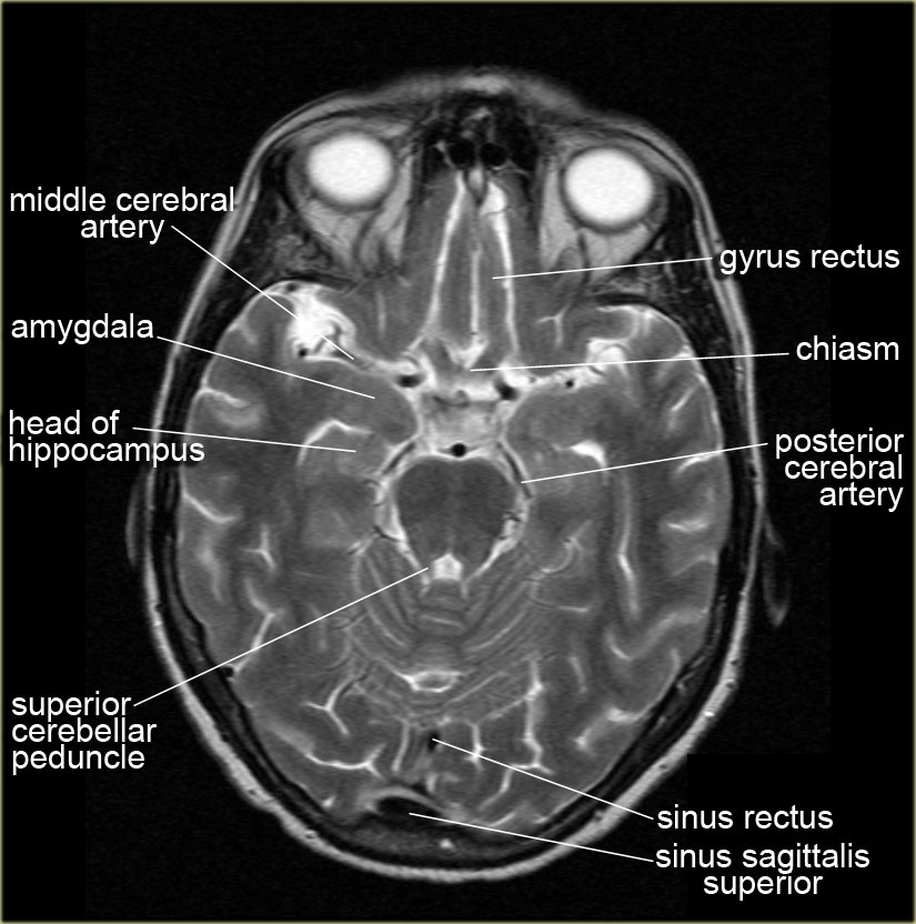
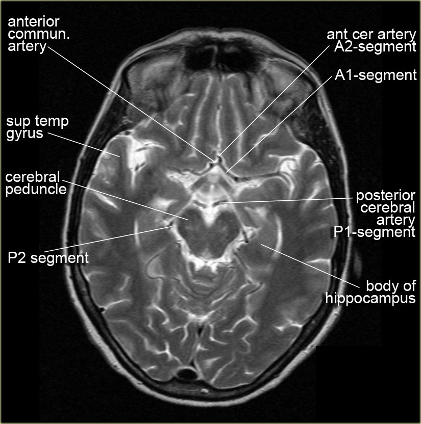
They lie on the ventricular surface of the hippocampus and become the fimbria of the fornix medially. Third and fourth ventricles in midline. Amygdala on ct and mr images the amygdala is a large region of gray matter contiguous with the uncus of the medial temporal lobe and the most anterior portion of the hippocampus the pes hippocampi.
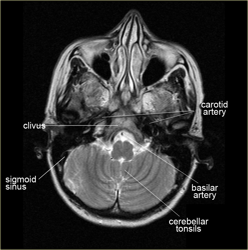
Real time interface human sectional anatomy. Ct of the chest lung windows axial anatomy. Brainstem and cerebellum without evidence of focal lesions.
These slides were taken from wikiradiography wetpaint here. Ct cross sectional anatomy of brain chest abdomen paranasal sinusus neck temporal bone heart slideshare uses cookies to improve functionality and performance and to provide you with relevant advertising. Focal abnormalities are not observed in the brain parenchyma.
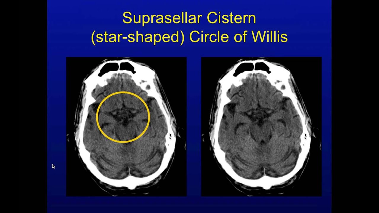
Radiological anatomy of the head and neck on a ct in axial coronal and sagittal sections and on a 3d images. Lateral ventricles of normal volume. Basal subarachnoid cisterns normal configuration.
Online mri ct sectional anatomy omcsa k anatomy is probably one of the most user friendly and convenient online interface for human anatomy atlas. Ct images of the brain are conventionally viewed from below as if looking up into the top of the head. Head ct anatomy normal anatomy 1.
Head and neck atlas.
 How To Interpret An Unenhanced Ct Brain Scan Part 1 Basic
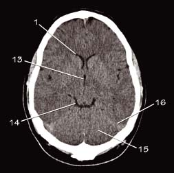
 Normal Ct Brain
 Radiology Basics Head Anatomy
 The Canine Head And Skull Ct Atlas Of Veterinary Clinical
Normal Brain Anatomy Ct And Mri Youtube
 Head Ct Anatomy Radiology Key
 The Radiology Assistant Brain Anatomy
 Headneckbrainspine
 Imaging Of Skull Base Pictorial Essay Raut Aa Naphade Ps
 Brain And Face Ct Interactive Anatomy Atlas
 Figure 69 5 From How To Read A Head Ct Scan Semantic Scholar
 Brain Imaging
 Ct Anatomy
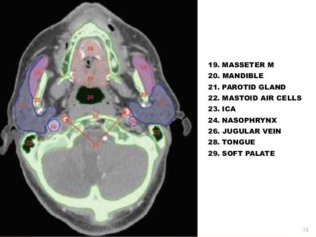 Ct Anatomy Head And Neck
 Head Ct Anatomy Radiology Key
 Normal Anatomy Of The Brain On Ct And Mri With A Few Normal
 Radiology Basics Head Anatomy
 Head Ct Interpretation Made Easy
 Brain And Face Ct Interactive Anatomy Atlas
 Head Ct
 The Radiology Assistant Brain Anatomy
 Head Ct
 The Radiology Assistant Brain Anatomy
 The Radiology Assistant Brain Anatomy
 Radiology Basics Head Anatomy
 Axial View Of A Head Computed Tomography Ct Scan Of Pineal
 Ct Anatomy
 Normal Head Ct
 Radiology Basics Head Anatomy
 Ct Scan Of Head And Neck
 Brain And Face Ct Interactive Anatomy Atlas
 Ct Scan Of Head And Neck
 Anatomy Of Orbit Bones Ct Google Search Ct Anatomy


Belum ada Komentar untuk "Ct Anatomy Of The Head"
Posting Komentar