Tragus Anatomy
An inner concentric ridge the antihelix surrounds the concha and. Because the tragus tends to be prominent in bats it is an important feature in identifying bats to species.
![]() Tragus Stock Vectors Royalty Free Tragus Illustrations
Tragus Stock Vectors Royalty Free Tragus Illustrations
The skin of the ear canal is very sensitive to pain and pressure.

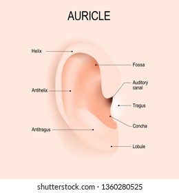
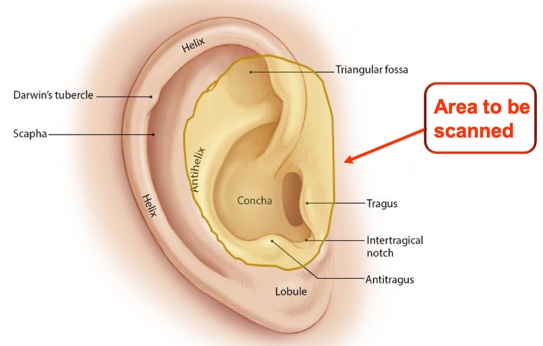
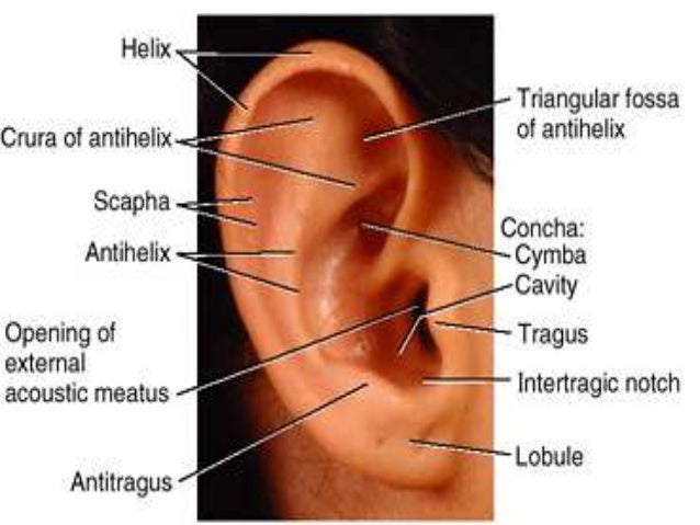
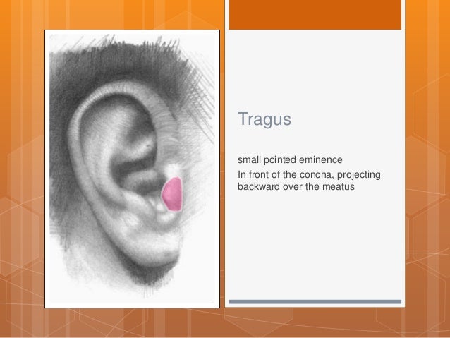
Tragus anatomy. Anatomy any of the hairs that grow just inside this entrance. There are three different parts to the outer ear. In front of the concha and projecting backward over the meatus is a small pointed eminence the tragus so called from its being generally covered on its under surface with a tuft of hair resembling a goats beard.
The tragus helix and the lobule. Prominence over and in front of the acoustic meatus. Hollow at the end of the helix.
As a piece of skin in front of the ear canal it plays an important role in directing sounds into the ear for prey location and navigation via echolocation. Anatomy head and neck areas tragus of the ear. Deep fossa of the external ear.
The canal is approximately an inch in length. Ear projection opposite the tragus. The tragus is a key feature in many bat species.
Fleshy part at the base of the ear. The tragus is a small cartilaginous structure and it is located on the anterior margin of the auditory canal. Outer ear two small projections the tonguelike tragus in front and the antitragus behind.
Variant anatomy of the external ear can be divided into congenital and acquired entities. Above the tragus a prominent ridge the helix arises from the floor of the concha and continues as the incurved rim of the upper portion of the auricle. Mouth of the canal of the temporal bone that carries sounds to the eardrum.
Congenital abnormalities of the ear are common and largely affect the shape of the auricle. Acquired entities can further be delineated into intrinsic processes such as cancer and extrinsic processes such as trauma. Anatomy the cartilaginous fleshy projection that partially covers the entrance to the external ear.
Ear canal the ear canal starts at the outer ear and ends at the ear drum. The outer ear is made up of cartilage and skin. Ear anatomy outer ear.
It is enclosed by special skin on its anterior and posterior parts and it is essential to the ear for its esthetic anatomical and functional aspects.
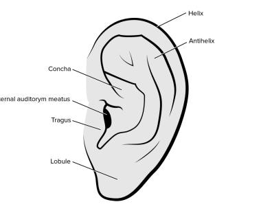 Ear Anesthesia Overview Indications Contraindications
 The Pinna Ear Anatomy Anatomy Tragus
Tragus Piercing Advanced Fundamentals Piercingnerd Com
Rbcp Earlobe Hypertrophy Correction
 A Anatomy B Measurements A 1 Helix Rim 2 Lobule 3
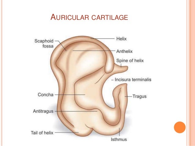 Anatomy Of External Ear And Middle Ear
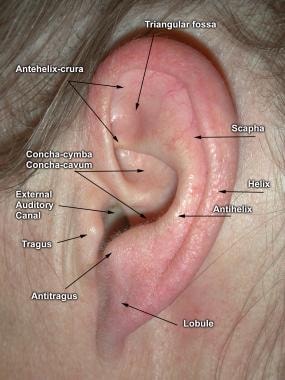 Dermatologic Approach To Ear Reconstruction Background
 Anatomy And Analysis Of The Ear Dr Shah
Ear Anatomy Prominent Ears Ear Pinning Surgery
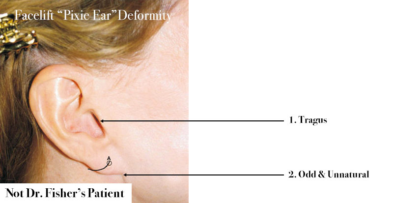 Minimal Scarring Best Facelift Beverly Hills
Tragus Piercing Advanced Fundamentals Piercingnerd Com
 Tragus Piercing Images Stock Photos Vectors Shutterstock
 Anatomical Landmarks Of Human Ear Download Scientific Diagram
Tragus Piercing Advanced Fundamentals Piercingnerd Com

 Cunningham S Text Book Of Anatomy Anatomy Fig 69
The Administration Of Ear Drops Cornerstone Ear Nose Throat
 Ear Auricular Anatomy Facial Anatomy Ear Parts Acupuncture
 Helix Triangular Fossa Superior Crus Inferior Crus Cymba

 External Anatomy Of The Ear Pinna Helix Antihelix Tragus
 The Ultimate Guide To Ear Piercings All Of The Piercings
 The External Ear Human Anatomy
 Otoscan 3d Ear Scanning The Future Is Now Jackie
 Related Image Ear Anatomy Ear Anatomy
 Pin By Shannon Gadsby On School Is Cool Ear Anatomy Human

Belum ada Komentar untuk "Tragus Anatomy"
Posting Komentar