Worm Internal Anatomy
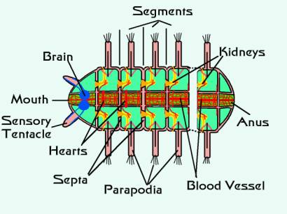
Internal anatomy of earthworm. Test your knowledge about anatomy of a common earthworm with this online quiz.
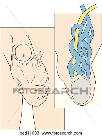 Male Genitalia Showing Anatomical Abnormality Of Left
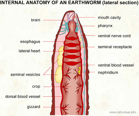
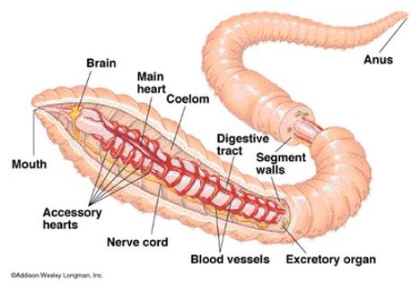
Internal anatomy of an earthworm lateral section.

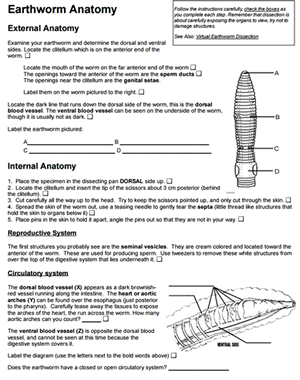
Worm internal anatomy. What is the internal anatomy of an earthworm. External dorsal surface of worm 1. Part of the digestive tract of an earthworm just after its mouth.
It is to be noted that the body of the earthworm is essentially a double tube. Insert the scissors into the opening and cut in a straight line all the way up through the mouth. Lay the worm on your dissecting tray with its dorsal side facing up.
They pull food into their mouths and then soak it in saliva. Use dissection pins to secure each end on the tray. Set of nerves in the abdomen of an earthworm.
The light colored tissue above and around the hearts are seminal vesicles. Lift up the skin with a pair of forceps and snip an opening with a pair of dissecting scissors. Small long cylindrical animal without legs or hard body parts.
The arrow points to the worms clitellum an organ responsible for mucus production during reproduction. Earthworm dissection picture guide. Underneath this in order are a thin layer of connective tissue a layer of circular muscle a layer of longitudinal muscle and a peritoneum surrounding the body cavity.
Earthworms push the pharynx from inside their mouths to grab hold of things. If a worm is cut open from the anterior to the posterior end by an incision through the body wall in the mid dorsal line the internal structures may easily be studied. Start your dissection about an inch posterior to the clitellum.
Internal earthworm anatomy. Entrance to the digestive tract of an earthworm. At the very front of an earthworm you will find the pharynx.
A trivia quiz called anatomy of a common earthworm. As a result segment 15 of one worm exudes sperm into segments 9 and 10 with its storage vesicles of its mate. One or more pairs of spermathecae are present in segments 9 and 10 depending on the species which are internal sacs that receive and store sperm from the other worm during copulation.
The dark line shows the location of the dorsal blood vessel. The worm is darker on its upper surface. Internal morphology overview 4 images.
Other reproductive parts appear as small white organs on the ventral side of the hearts. General anatomy of a polychaete the outer surface of the body wall consists of a simple columnar epithelium covered by a thin cuticle. Highscores 207 registered players.
 Hemichordates
 Polychaete Wikipedia
 Earthworm A Detailed Look Anyone From A Student To A
 Polychaete Annelid Identification Or You Can Always Tell
 Earthworm Wikipedia
 Earthworm Dissection
 Worm Lab
 Earthworm Anatomy Key
 Worms Lessons Tes Teach
 Worms
Worms
 Internal Anatomy Earthworm Lateral Visual Dictionary
 Worm Dissection 2014 Warning Stay Focused In This Lab Ppt
 Internal Anatomy Of A Male C Elegans Nematode Marine
 53 Lumbricus Terrestris Lumbricidae A External Features
 Worm Anatomy Physiology Compost Ology City Of Euless
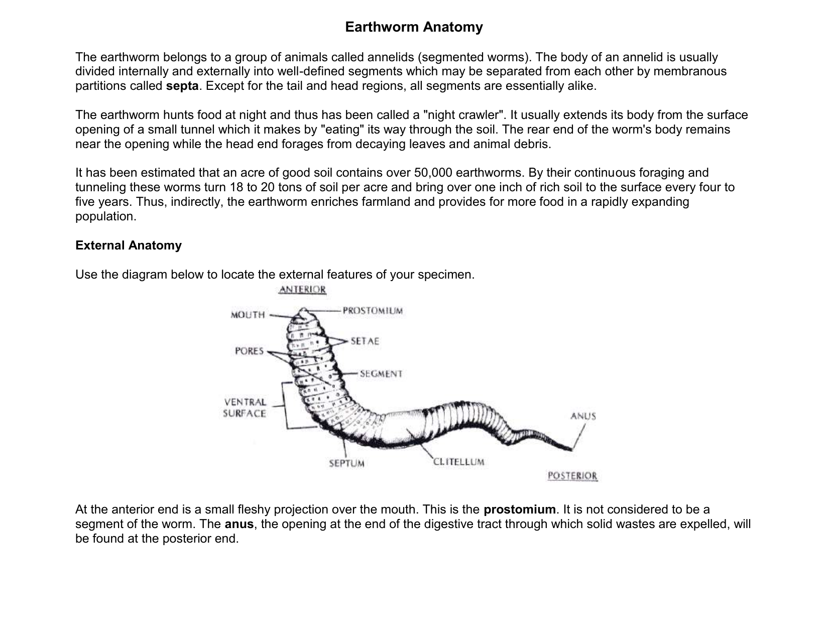
 Earthworm Anatomy
 Anatomy Of The Earthworm Midlakes Pages 1 4 Text
 Earthworms
 Phylum Hemichordata Pterobranchs And Acorn Worms Amphibians
 Earthworm Anatomy
 Anatomy Wormwatch
 Earthworm Anatomy Anatomy Project
 Earthworm Anatomy And Dissection Guide Biology Junction
 Red Worm Anatomy Eisenia Fetida The Perfect Composting Worm
 Annelida


Belum ada Komentar untuk "Worm Internal Anatomy"
Posting Komentar