Fossa Anatomy
The fossa of rosenmüller also known as the posterolateral pharyngeal recess is the most common site of origin for nasopharyngeal carcinoma. The popliteal fossa is 25 cm wide and mainly consists of fat tissue.
 Pterygopalatine Fossa Wikipedia
The fossa appears as a diminutive form of a large felid such as a cougar but with a slender body and muscular limbs and a tail nearly as long as the rest of the body.

Fossa anatomy. It is the main path by which vessels and nerves pass between the thigh and the leg. The median cubital vein joins the two longest vessels that run up the length of your arm. Glenoid cavity glenoid fossa the concavity in the head of the scapula that receives the head of the humerus to form the shoulder joint.
Cerebral fossa any of the depressions on the floor of the cranial cavity. Fossa a concavity in a surface especially an anatomical depression pit. It has a mongoose like head relatively longer than that of a cat although with a muzzle that is broad and short and with large but rounded ears.
Gross anatomy it is located superior and posterior to the torus tubarius the posterior projection of. The antecubital fossa is the shallow depression located in front of the median cubital vein of your arm. The superomedial aspect of the popliteal fossa is bounded by the semimembranosus and.
The cubital fossa is triangular in shape and thus has three borders. Condylar fossa condyloid fossa either of two pits on the lateral portion of the occipital bone. Blood vessels are located deep to the nerves within the fossa and include.
A trench or channel. Lateral border medial border of the brachioradialis muscle. The popliteal fossa is a diamond shaped area located on the posterior aspect of the knee.
Amygdaloid fossa the depression in which the tonsil is lodged. Superior border hypothetical line between the epicondyles of the humerus. Medial border lateral border of the pronator teres muscle.
In anatomy a hollow or depressed area. In this article we shall look at the anatomy of the popliteal fossa its borders contents and clinical correlations. The temporal fossa is a shallow depression on the temporal lines and one of the be massive marks on the skull.
The occipital bones including temporal bone sphenoid bone parietal bone and the frontal bone put up to its concave wall.
Cubital Fossa Radiology Reference Article Radiopaedia Org
 7 Tmj Infratemporal Fossa Anatomy Physiology Spom
![]() Temporal Fossa Anatomy Borders And Contents Kenhub
Temporal Fossa Anatomy Borders And Contents Kenhub
 Infratemporal Fossa Wikipedia
 Middle Cranial Fossa Surgery Offers A Better Chance For
 Cubital Fossa Wikipedia
 Anatomy Of The Posterior Fossa Clinical Gate
 Figure Cubital Fossa Image Courtesy S Bhimji Md
 Temporal Fossa Anatomy Qa
 Fossa Anatomy Anatomy System Human Body Anatomy Diagram
 Head And Neck Anatomy 11
 The Anatomy Of The Posterior Cranial Fossa Springerlink
 Middle Cranial Fossa Boundaries Contents Teachmeanatomy
 Ischio Rectal Ischio Anal Fossa Anatomy Qa
 Anterior Cranial Fossa
 Lacrimal Bone Anatomy
 3d Printed Deep Face Infratemporal Fossa Model
 Posterior Cranial Fossa Boundaries Contents Teachmeanatomy
 Anatomy Infratemporal Fossa Flashcards Quizlet
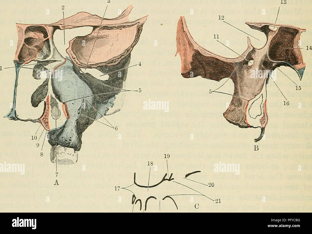 Cunningham S Text Book Of Anatomy Anatomy 170 Osteology
 Popliteal Fossa Knee Joint Anatomy Medical Anatomy
 A Study Of Middle Cranial Fossa Anatomy And Anatomic
 The Shoulder Bony Anatomy Part 1 Tami Apland
 Openings In The Cranial Fossa Skull Anatomy Sphenoid Bone


Belum ada Komentar untuk "Fossa Anatomy"
Posting Komentar