Triad Anatomy
Found in walls of large arteries in large airways of lungs in arrector pili muscles and in radial and circular muscles of iris when the stimulus comes it only stimulates one or two. See portal triad for discussion of misnomer mouseover for labeled slide references.
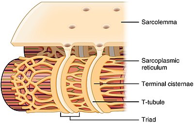
Each skeletal muscle fiber has many thousands of triads visible in muscle fibers that have been sectioned longitudinally.

Triad anatomy. The number of triad s per sarcomere depends on the species. In the histology of skeletal muscle a triad is the structure formed by a t tubule with a sarcoplasmic reticulum sr known as the terminal cisterna on either side. A group of three similar bodies or a complex composed of three items or units.
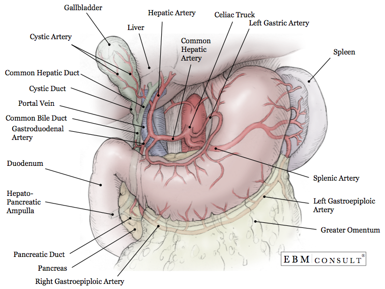
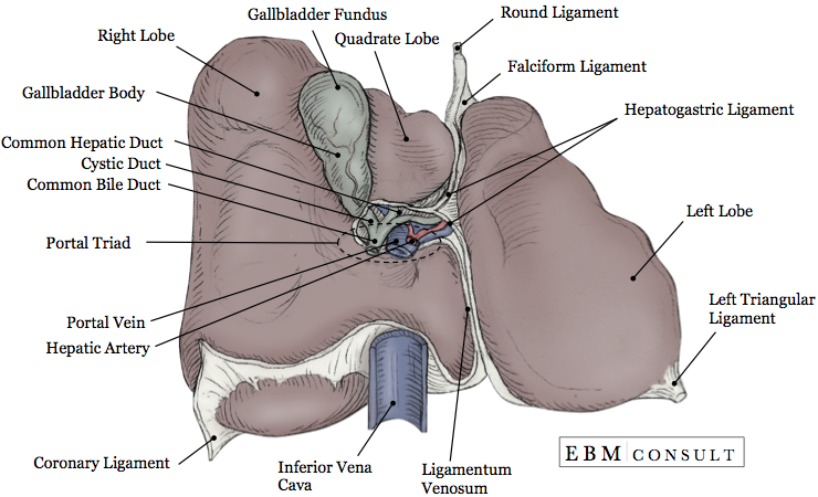
It can refer both to the largest branch of each of these vessels running inside the hepatoduodenal ligament and to the smaller branches of these vessels inside the liver. Stimulation causes contraction of only one fiber. This would require urgent evaluation by a trained physician.
Individual fibers each with own motor neuron terminals and with few gap junctions between neighboring fibers. An element with a valence of three. A portal triad is an arrangement in the liver which consists of a hepatic artery a portal vein and a bile duct.
Treatment of the unhappy triad depends on the severity of the injury and the extent of damage to the surrounding structures. The unhappy triad labelled in complete anatomy. For example in frog muscle there is one per triad and in mammalian muscle there are two.
In fishes and crustaceans only one cisterna is associated with each transverse tubule thus forming a dyad. In the smaller portal triads the four vessels lie in a network of connective tissue and are surrounded on all sides by hepatocytes. If surgery is required most surgeries are done using a minimally invasive approach called arthroscopy.
A three element complex called a triad. Odonoghue unhappy triad or terrible triad often occurs in contact sports such as basketball football or rugby when there is a lateral force applied to the knee while the foot is fixated on the groundthis produces the pivot shift mechanism. The odonoghue unhappy triad comprises three types of soft tissue injury that frequently tend to occur simultaneously in knee injuries.
A larger one that arises from the clavicle the sternum the cartilages of most or all of the ribs and the aponeurosis of the external oblique muscle and is inserted by a strong flat tendon into the posterior bicipital ridge of the humerus. Triad anatomy jump to navigation jump to search. Ts of the tarsus.
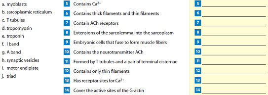
The triad allows an electrical impulse traveling along a t tubule to stimulate the membranes of adjacent sacs of the sr describe the structure of thin and thick myofilaments and name the kinds of proteins that compose them. The various combinations of usually three injuries that occur in trauma to the hock joint based first on injury to the central tarsal bone.
 Anat20006 Lecture Notes Winter 2017 Lecture 30 Cystic
Hepatoduodenal Ligament Wikipedia
 Anatomy Definition And Treatment Of The Terrible Triad Of
 The Unhappy Triad Sparcc Sports Medicine Tucson Az
Heart Valve Repair Or Replacement Triad Cardiac And
 Unhappy Triad Or Blown Knee Or Terrible Triad Etiology
 Portal Triad Iphone Cases Fine Art America
Sean Lee And The Unhappy Triad In Street Clothes
 Surreal Portal Triad Of The Liver Art In Anatomy
 Liver Anatomy A Entire Organ And Blood Supply Blue

 Anatomy And Physiology Of The Liver
 Liver Blood Vessel An Overview Sciencedirect Topics
 Unique Portal Triad Art Fine Art America
 View Large Accessmedicine Mcgraw Hill Medical
 Triad Organization In Skeletal Muscle Left Electron
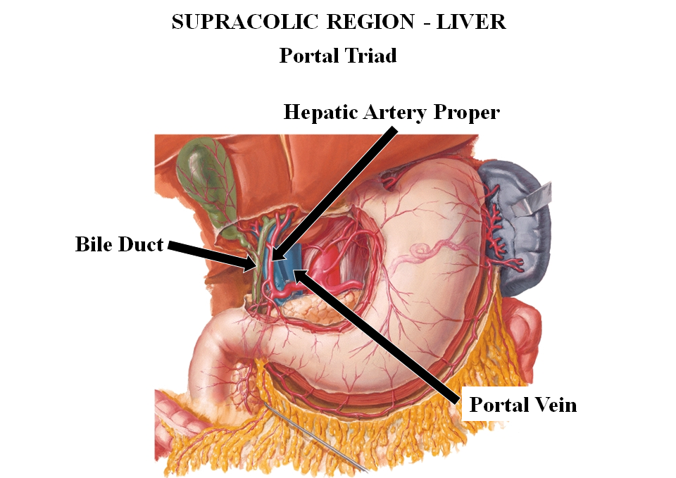
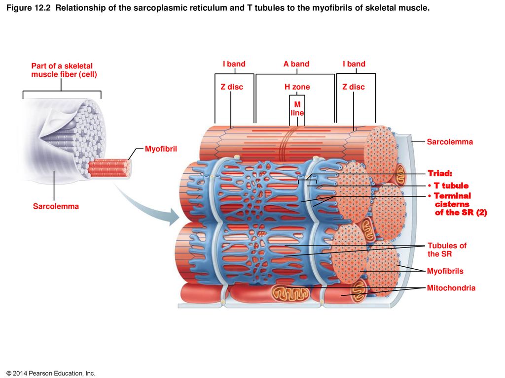 Figure 12 1 Microscopic Anatomy Of Skeletal Muscle Ppt
 Structure And Function Of The Triad A The Triad Is A
Skeletal Muscle Anatomy And Physiology Openstax
 Chapter 9 Solutions Visual Anatomy Physiology 2nd
Anatomy Physiology Muscle And Muscle Tissue 13 10

Belum ada Komentar untuk "Triad Anatomy"
Posting Komentar