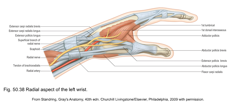
Anatomy Of The Left Wrist
The wrist connects the hand to the forearm. Profundus tendons which pass through the palm side of the wrist and hand.
 Wrist Hand Anatomy
It is relatively prone to injury.

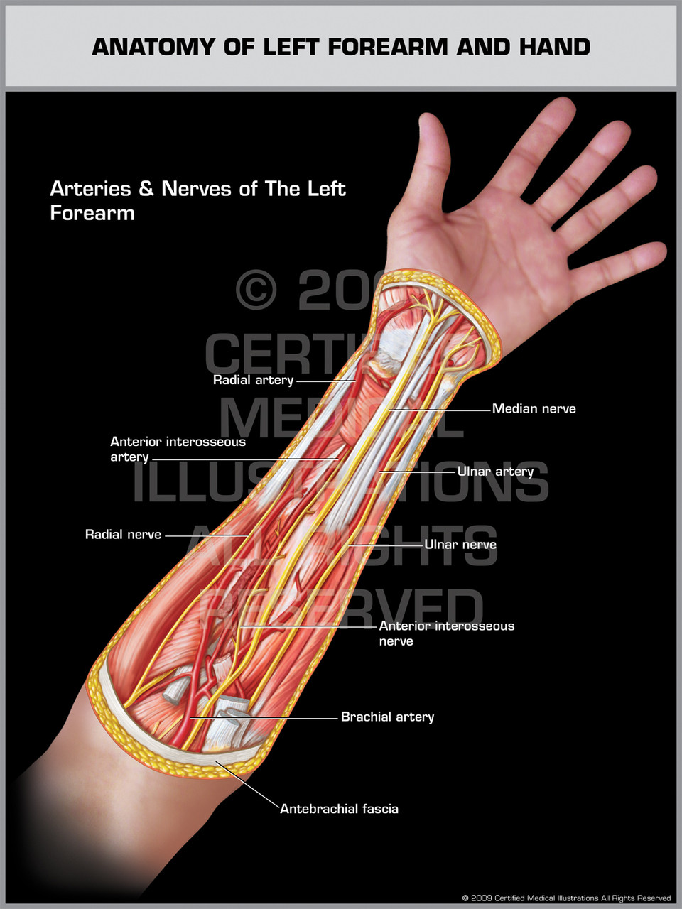
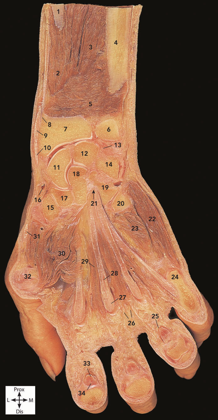
Anatomy of the left wrist. The exhibit depicts the anatomy of the left forearm featuring arteries and nerves. Superficialis tendons which pass through the palm side of the wrist and hand. Anatomy and injuries of the hand and wrist illustrates the following normal anatomy.
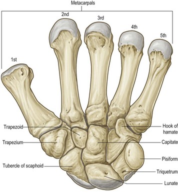
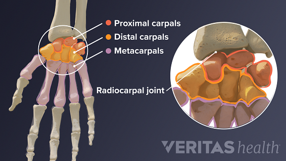
The carpal bones are arranged in 2 interrelated rows. These bones also form the flexible wrist joint with the proximal row of the carpals. One row connects with the ends of the bones in the forearm radius and ulna.
Rotation of the radius around the ulna results in the supination and pronation of the hand. 2 the wrist joint or radiocarpal joint the joint between the radius and the carpus and 3 the anatomical region surrounding the carpus including the distal parts of the bones of the. There are two long bones in the forearm that run from the elbow to the wrist.
Anatomy of the hand and wrist. The wrist must be extremely mobile to give our hands a full range of motion. The wrist can bend straighten move laterally and rotate.
The anatomy of the wrist joint is extremely complex probably the most complex of all the joints in the body. The main tendons of the hand are. The smaller bone the ulna is on the little finger side.
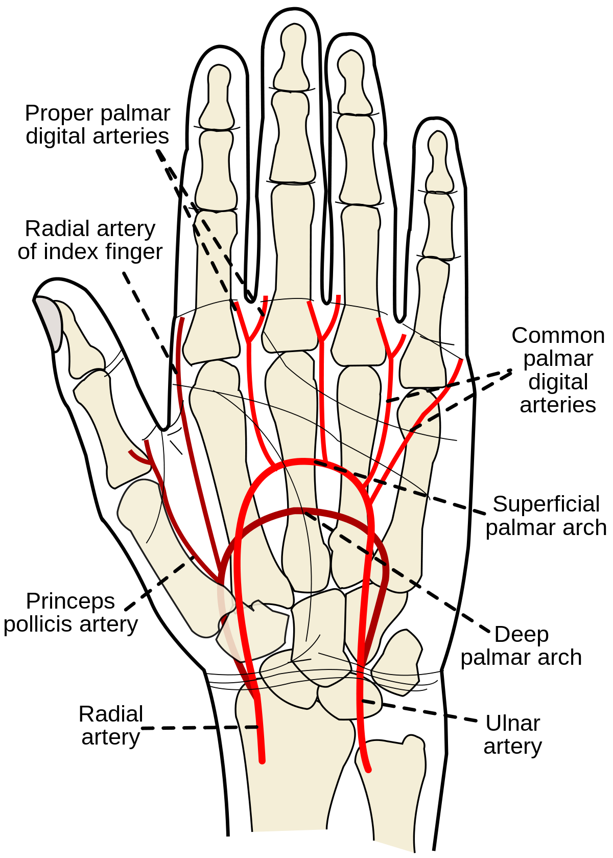
The larger bone the radius is on the same side as the thumb. Please call 1 888 999 0410 for custom sizing and other options. The wrist is a complex mechanical system of 8 small bones known as the carpal bones.
The wrist is actually a collection of many bones and joints. The forearms ulna and radius support the many muscles that manipulate the bones of the hand and wrist. It consists of the distal ends of the radius and ulna bones eight carpal bones and the proximal ends of five metacarpal bones.
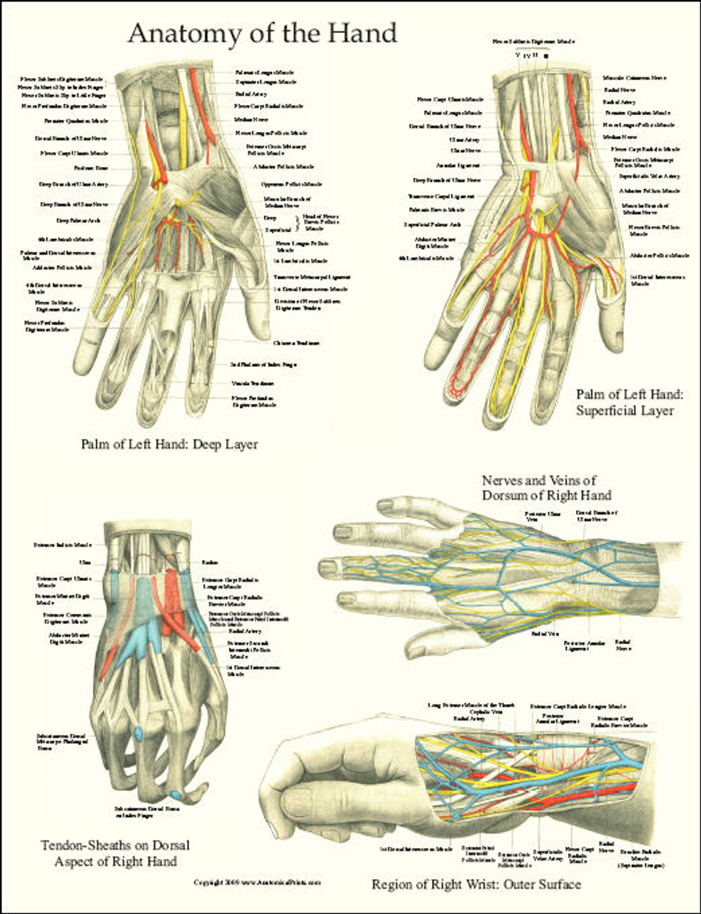
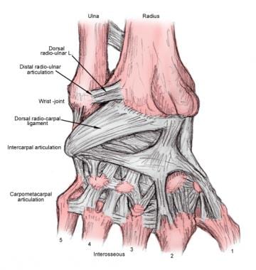
These bones and joints let us use our hands in lots of different ways. Extensor tendons of the fingers which attach to the middle and distal phalanges and extend. Palmar view of the bones mucsles and ligaments of the hand and wrist dorsal view of the bones muscles and ligaments of the foot and ankle nerve and blood supply to the hand and wrist deep dorsal view of the left wrist deep volar view of the left wrist.
This arrangement of bones allows for a wide range of movement. Bones muscles tendons nerves. The joints of the wrist are surrounded by a fibrous capsule and are held together by an array of ligaments that provide carpal stability by linking the bones both dorsally and volarly see the.
In human anatomy the wrist is variously defined as 1 the carpus or carpal bones the complex of eight bones forming the proximal skeletal segment of the hand. The wrist links the hand to the arm.
 Research Assistant Resource Update Access Medicine Ucla
 Applied Anatomy Of The Wrist Thumb And Hand
 Wrist Hand Atlas Of Anatomy
 Median Nerve Wikipedia
 Left Wrist Anatomy Medical Illustration Human Anatomy
 Right And Left Wrist Injuries High Impact Visual
 Hand And Wrist Anatomical Chart
The Upper Limbs Human Anatomy And Physiology Lab Bsb 141
 Left Hand And Wrist Bones Labeled On White Background Stock
 Carpal Tunnel Syndrome Symptoms And Treatment Orthoinfo
 Wrist Anatomy Eorthopod Com
Carpal Tunnel Syndrome Cleveland Clinic
 An Anatomical Picture Of The Left Forearm The Brachial
 Anatomy Of Left Forearm Hand
 Hand And Wrist Anatomy Laminated Poster
 Radial Artery Wikipedia
 Logan S Illustrated Human Anatomy
 Left Hand Tendons Diagram Reading Industrial Wiring Diagrams
 Wrist Joint Anatomy Overview Gross Anatomy Natural Variants
 Wrist Anatomy Eorthopod Com
 Normal Anatomy Vs Left Wrist Fracture Trial Exhibit
 Left Wrist Fracture Medical Illustration Human Anatomy
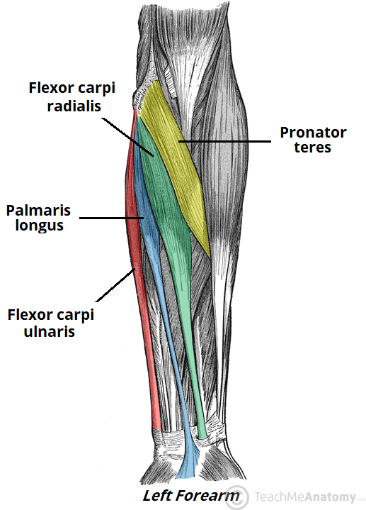 Muscles Of The Anterior Forearm Flexion Pronation
 Anatomy Of The Wrist Thumb And Hand Musculoskeletal Key
 Forearm Pain Relief Cause And Treatment Deep Recovery
 Guide To Wrist Anatomy


Belum ada Komentar untuk "Anatomy Of The Left Wrist"
Posting Komentar