Anatomy Sonography
The following fetal parts are checked during the anatomy ultrasound. Oblique left showing the ligamentum teres.
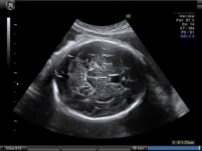
 Central Nervous System Diagnosis Of Fetal Abnormalities
When a level 2 ultrasound is done.

Anatomy sonography. I wasnt ready for how intense my 20 week anatomy scan was. By the end of my pregnancy. Neck nuchal fold thickness.
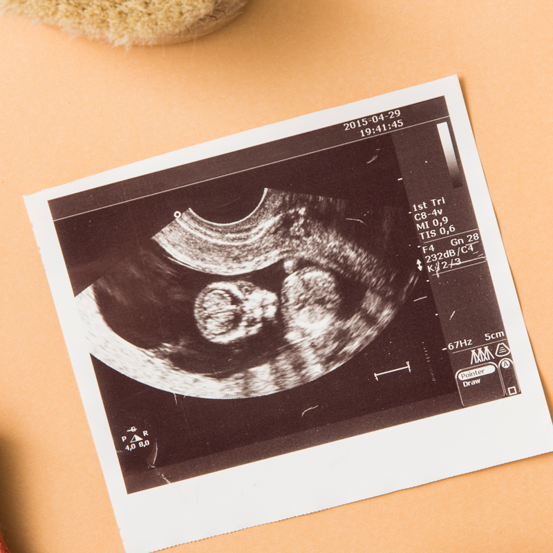
Most anatomy scans are performed in the second trimester of pregnancy typically at 20 weeks but they can be done anytime between 18 weeks and 22 weeks. Can you give us a brief description of what the 20 week anatomy scan is. The scan is performed transabdominally.
In short its a scan where we can get the most information about your babys anatomy. Those who want to can find out the sex of the baby if desired. The second trimester extends from 13 weeks and 0 days to 27 weeks and 6 days of gestation although the majority of these studies are performed between 18 and 23 weeks.
Its done right in the middle of the pregnancy because at that point most babies are large enough that you can see all of the structures that you need to see. If you have a condition that needs to be monitored such as carrying multiples you may have more than one detailed ultrasound. Find out what youll see when you have yours.
Segmental anatomy according to couinaud. The anatomy scan is a level 2 ultrasound which is typically performed on pregnant women between 18 and 22 weeks. In women at high risk for preterm delivery multiple pregnancies previous preterm birth abnormalities of the uterus or previous cervical surgery we may also carry out a transvaginal scan to measure the length of the cervix.
The ligamentum venosum is highlighted in orange. The second trimester scan is a routine ultrasound examination in many countries that is primarily used to assess fetal anatomy and detect the presence of any fetal anomalies. Choose from 500 different sets of ultrasound sonography anatomy flashcards on quizlet.
Brain ventricles choroid plexus mid brain posterior fossa cerebellum cisterna magna. Heart rate rhythm 4 chamber views. Hover over the images for highlighted anatomy.
Learn ultrasound sonography anatomy with free interactive flashcards. Was that i got a sneak peak at my babies with an ultrasound every time i went to the doctor for a checkup. The 20 week ultrasound or anatomy scan is an eagerly anticipated ultrasound for parents.
This is a detailed scan of your babybabies anatomy. Porta hepatis is seen with an oblique angle 45degree rotation from the sagittal view to the transverse view. Skull shape integrity bpd and hc measurements.
 Anatomy And Sonography Of The Abdominal Vasculature Diagram
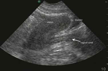 Pelvic Ultrasound Chapter 7 Atlas Of Emergency Ultrasound
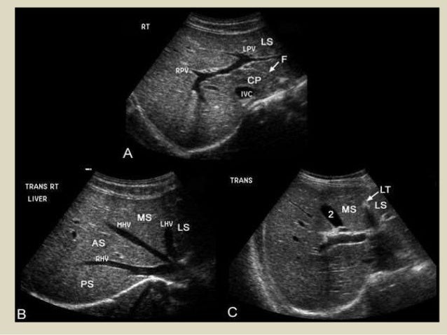
 Liver Anatomy And Segments By Ultrasound In Arabic
Figure 1 From Chest Wall Underappreciated Structure In
 20 Week Ultrasound Anatomy Scan Lani
 Imaging Anatomy Ultrasound 9780323548007 Medicine
 Ultrasound Of Liver Segments Anatomy
 The Anatomy Scan Ultrasound Is An Amazing Experience You Ll
 Measurement In Ultrasound
 Free Chapter Normal Cns Ultrasound Brain Anatomy Ob Images
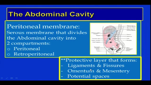 Abdominal Sonography Anatomy Overview And Scan Fundamentals
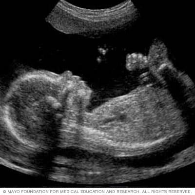 Fetal Ultrasound Mayo Clinic
 Mickey Mouse Sign Portal Vein Cbd Ha Ultrasound
 Ultrasound Images Of Fetal Brain
 Introduction To Renal Ultrasound Ultrasound Physics
 Breast Anatomy The Basis For Understanding Sonography
 Presentation1 Abdominal Ultrasound Anatomy

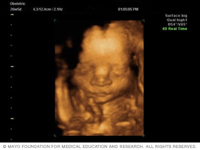 Fetal Ultrasound Mayo Clinic
 Porta Hepatis Lymph Node Liver Anatomy Sonography

 1 Normal Ultrasound Anatomy Seen Above The Asis Download
 Normal Pancreas Ultrasound Radiology Case Radiopaedia Org
 Heart Anatomy The Cardiovascular System Circulatory System
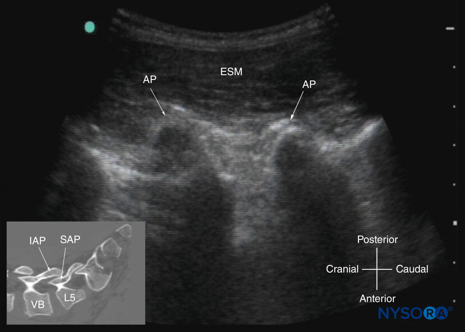 Spinal Sonography And Applications Of Ultrasound For Central
 Sonography Aorta Cranially Heart Celiac Branch Sagittal
 Video Normal Shoulder Scanning Technique Sonography
 Musculoskeletal Sonography Technique Anatomy Semeiotics And Pathological Findings In Rheumatic Diseases
 Sonographic Anatomy Of Female Pelvis Simplified Approach

 Liver Hilum Anatomy Ultrasound
 Sonography Introduction To Normal Structure And Function
Belum ada Komentar untuk "Anatomy Sonography"
Posting Komentar