Hip Anatomy Nerves
The nerve signals carried by the femoral nerve are a critical part of the ability to stand walk and maintain balance. A smaller nerve called the obturator nerve also goes to the hip.
 Ultrasound Guided Obturator Nerve Block Nysora
Anatomy nerves are complex structures that branch out like a tree.
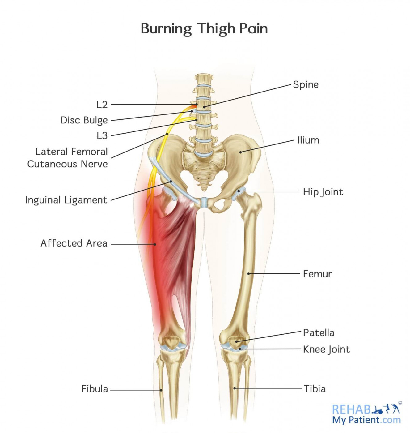
Hip anatomy nerves. The main nerves are the femoral nerve in front and the sciatic nerve in back of the hip. All of the nerves that travel down the thigh pass by the hip. Three nerves run through the region of the anterior and medial thigh.
The obturator nerve in addition to supplying the hip is responsible for thigh sensation. Hip joint capsule attaches anteriorly to the along the intertrochanteric crest. This joint allows a wide range of movements of the lower limbs and is used when walking running climbing lunging and bending.
Three ligaments iliofemoral ligament y ligament of bigelow strongest ligament. Extends posteriorly only partially across the femoral neck basicervical and intertrochanteric regions are extracapsular. Shakira and justin just use the hip and thigh anatomy to its full potential.
The main nerves of the hip that supply the muscles in the hip include the femoral obturator and sciatic nerves. If you have a pinched nerve in your hip walking. Clinical anatomy for dummies.
A pinched nerve in the hip often causes pain in the groin. Anatomy of the hip. This nerve runs from the lumbar plexus along the psoas major past the inguinal ligament to enter the femoral triangle.
Sometimes the pain also radiates down the inner thigh. In vertebrate anatomy hip or coxa in medical terminology refers to either an anatomical region or a joint. Aiis to intertrochanteric line.
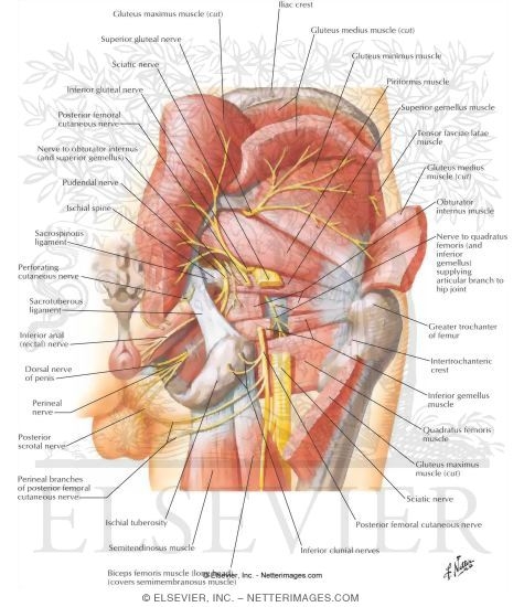
The nerves in the hip include the obturator nerve the lateral femoral cutaneous nerve the femoral nerve and the sciatic nerve. The sciatic nerve is the most commonly recognized nerve in the hip and thigh. The most recognizednotable nerve in the thigh and hip is probably the sciatic nerve.
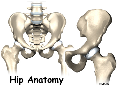
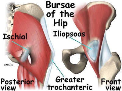
It has branches that innervate the anterior thigh muscles and the hip joint. The hip is a major ball and socket joint connecting the long bones of the lower limbs femur to the pelvis. The hip region is located lateral and anterior to the gluteal region inferior to the iliac crest and overlying the greater trochanter of the femur or thigh bone.
In this page we will focus on the anatomy of the hip and thigh and discover the incredible functions of this part of the human body. The sciatic nerve is largeas big around as your thumband travels beneath the gluteus maximus down the back of the thigh where it branches to supply the muscles of the leg and foot. It can travel to the knee as well.
These nerves carry the signals from the brain to the muscles that move the hip.
 Imaging Of Neuropathies About The Hip Martinoli Ultrasound
 Muscles Advanced Anatomy 2nd Ed
Anterior Hip Pain Lesser Known Causes Transform
 Femoral Nerve Anatomy Pictures And Information
 Hip Clinical Gate Nerve Anatomy Muscle Nerve Leg Anatomy
 Anatomy And Injuries Of The Hip Anatomical Chart
 Muscles Advanced Anatomy 2nd Ed
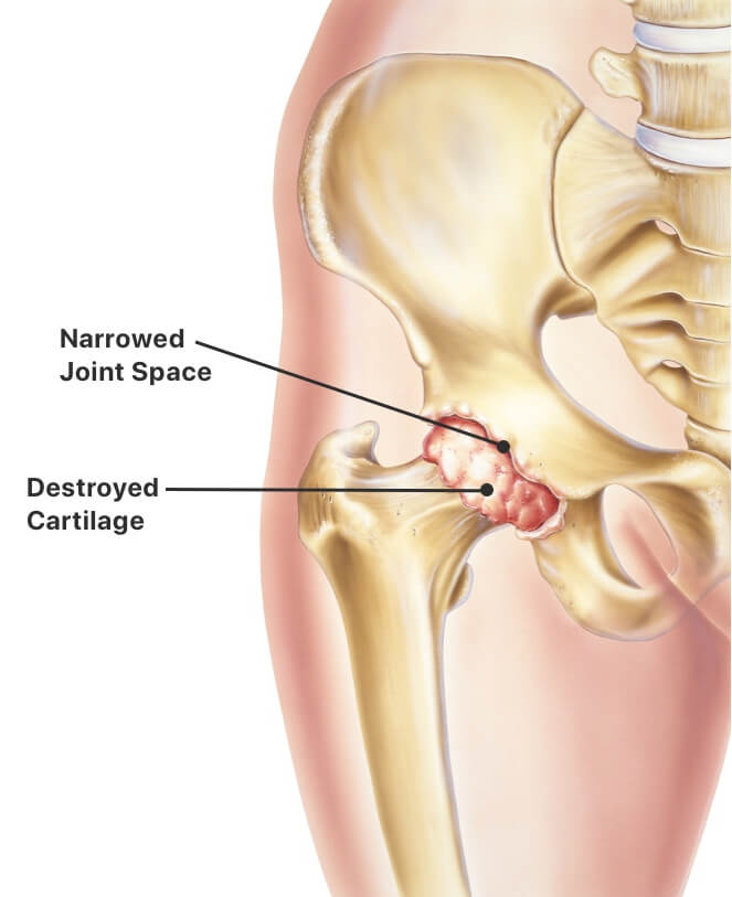 Hip Replacement Procedure Types Recovery Time And Risks
 Anatomy And Injuries Of The Hip Anatomical Chart
 Hip Joint W Sciatic Nerve Model Human Body Anatomy Replica Of Normal Muscled Hip Joint W Sciatic Nerve For Doctors Office Educational Tool Gpi
 Nerves Of The Leg And Foot Interactive Anatomy Guide
 Burning Thigh Pain Rehab My Patient
:watermark(/images/watermark_5000_10percent.png,0,0,0):watermark(/images/logo_url.png,-10,-10,0):format(jpeg)/images/atlas_overview_image/723/rhPG1aJnnONltuWiHn0awA_nerves-vessels-pelvis-thigh_english.jpg) Diagram Pictures Neurovasculature Of The Hip And The
Diagram Pictures Neurovasculature Of The Hip And The
:background_color(FFFFFF):format(jpeg)/images/library/12447/nerves-of-female-pelvis_english.jpg) Pelvic Veins Lymphatics And Nerves Anatomy And Drainage
Pelvic Veins Lymphatics And Nerves Anatomy And Drainage
 Surface Models Of The Hip Joint Muscles And Their
:background_color(FFFFFF):format(jpeg)/images/library/11030/Hip_and_thigh_1.png) Hip And Thigh Bones Joints Muscles Kenhub
Hip And Thigh Bones Joints Muscles Kenhub




Belum ada Komentar untuk "Hip Anatomy Nerves"
Posting Komentar