Anatomy Knee Bursa
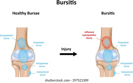
Knee bursitis causes pain and can limit your mobility. Anatomy of the knee bursae a bursa is a small sac made of fibrous tissue that has an inner lining of synovial type membrane.
 A Medical Illustration Of A Knee With An Inflamed Prepatellar
The knee contains three important groups of bursae.

Anatomy knee bursa. So lets have a look at knee bursitis anatomy particularly focusing on the 5 main knee bursa which are the ones that are most commonly injured. A knee bursa also known as a subcutaneous prepatellar bursa aids with movement when we walk run stretch or even cross our legs. Between the patellar ligament and the anterior part of the tibia bursitis of the knee is an inflammation of any of those eight bursae resulting in pain and tenderness of the knee swelling and a warm feeling when you touch the area.
The prepatellar bursae lie in front of the patella. It protects the patella. The pcl is in the center of the knee it limits backward leg movements.
This is usually when there is excessive friction over the bursa causing it to either become inflamed or when it dries out so it no longer works properly. Bursae one is a bursa are fluid filled sacs that help cushion the knee. The prepatellar bursa is one of the larger bursae of the knee and is located on the front of the patella hence pre patellar just under the skin.
The knee bursae are the fluid filled sacs and synovial pockets that surround and sometimes communicate with the knee joint cavity. Knee bursae are sacs surrounding the knee joint that are filled with synovial fluid. The knee bursae can be either communicating or non communicating with the knee joint itself.
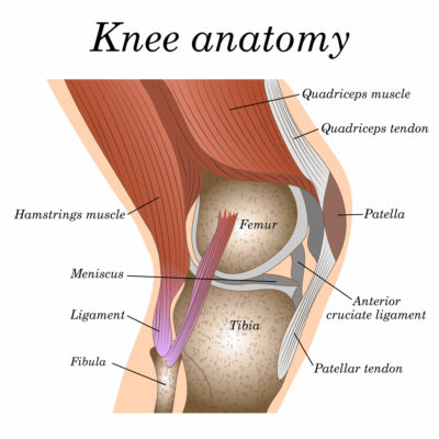
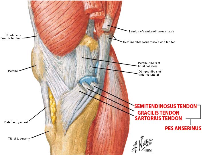
The bursae can become irritated by frequent kneeling. They represent the weak point of the joint but also provide enlargements to the joint space. Knee tendons and ligaments.
A knee bursa basically functions as a cushion. The mcl runs along the inside of the knee joint it provides stability to the medial inner part of the knee. There are bursa located underneath the tendons and ligaments on both the lateral and medial sides of the knee.
When one becomes inflamed increased tension and pain can occur in a temporary condition known as bursitis. Knee bursitis is inflammation of a small fluid filled sac bursa situated near your knee joint. The acl is in the center of the knee it limits rotation and forward leg movements.
It is filled with synovial fluid or lubricant made by the membrane. The bursae are thin walled and filled with synovial fluid. They facilitate movement and reduce friction where tendons or muscles pass over bony prominences.
 Pain Around The Knee Bursitis Jonathan Aarons Md Pain
 Knee Bursitis Information Sinew Therapeutics
 Bursitis Of The Knee Healthlink Bc
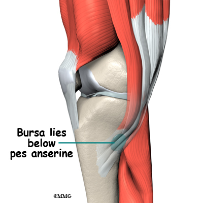 Pes Anserine Bursitis Of The Knee Eorthopod Com
 Knee Bursitis Information Sinew Therapeutics
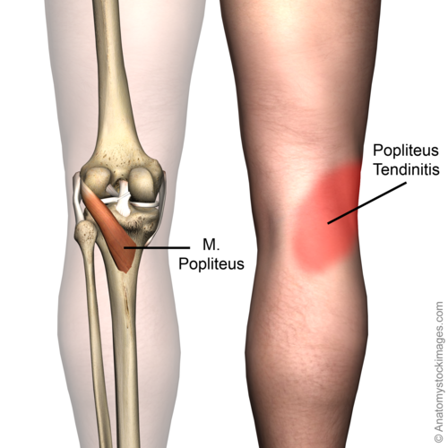 Popliteus Tendinopathy Physiopedia
 Bursae Knee Pain Inflamed Ruptured Bursa Sac

 Prepatellar Bursitis

 What Causes A Swollen Knee Water On The Knee
 Tiny Bursa Balloons To Bursitis Direct Orthopedic Care
 Knee Bursitis Prepatellar Bursitis Everything You Need To Know Dr Nabil Ebraheim
Elbow Olecranon Bursitis Orthoinfo Aaos
Bursitis Ankle Bursa Care And Prevention
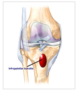 Infrapatellar Bursitis Natural Treatment Osmo Patch Us
Hip Bursitis Orthoinfo Aaos
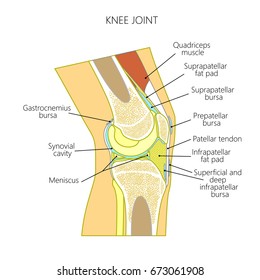
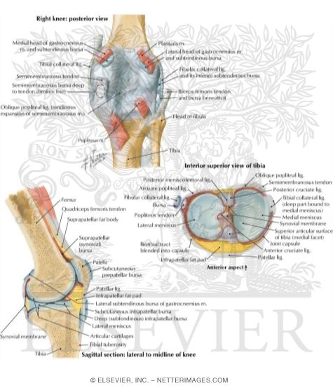 Knee Joint Ligaments And Bursae
 Bursitis Practice Essentials Anatomy Pathophysiology
 Synovial Bursa Wikipedia
 Bursitis Knee
 Knee Bursa Images Stock Photos Vectors Shutterstock
 Knee Bursitis Images Stock Photos Vectors Shutterstock
 Pes Anserinus Bursitis Tendinopathy
 Pes Anserinus Bursitis Symptoms And Treatment Bone And Spine
 Prepatellar Bursitis Orthopedic Knee Specialist Richmond Va
Belum ada Komentar untuk "Anatomy Knee Bursa"
Posting Komentar