Extensor Tendon Anatomy
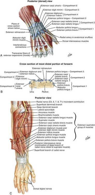
The common extensor tendon is a tendon that attaches to the lateral epicondyle of the humerus. It contains two tendons the abductor pollicis longus abd l and the extensor pollicis brevis est b both of which originate deep in the ulnar side of the forearm from the dorsal aspects middle third of the ulna radius and interosseous membrane b.
 Forearm Pain Relief Cause And Treatment Deep Recovery
It is however commonly brought on by activities that require repetitive wrist flexion and extension.

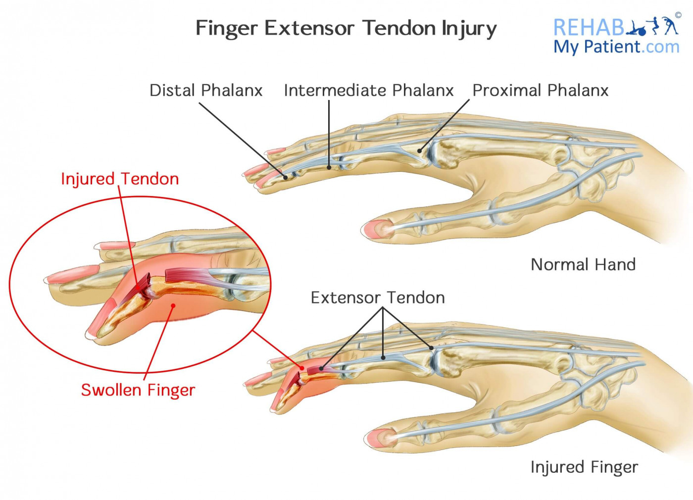
Extensor tendon anatomy. Any movement of the thumb and wrist causes the patient pain inflammation and swelling. They can be injured by a minor cut or jamming a finger which may cause the thin tendons to rip from their attachment to bone. They lie next to the bone on the back of the hands and fingers and straighten the wrist fingers and thumb figure 1.
These particular tendons allow you to straighten your fingers and thumb and can be injured by a simple cut or jammed finger. Intersection syndrome can be caused by direct trauma to the second extensor compartment. And vilensky j all in one anatomy exam review.
Extensor pollicis brevis epb. Indications lacerations 50 of tendon in all zones if patient can extend digit against resistance. Extensor tendons are just under the skin.
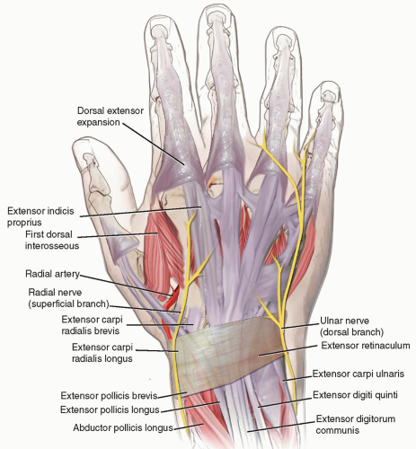
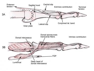
Each tunnel is lined internally by a synovial sheath and separated from one another by a fibrous septa. Understanding of and familiarity with the extensor anatomy of the hand and fingers by the radiologist is crucial for better assessment of pathologic conditions with mr imaging and optimization of this mo dality as a diagnostic tool. The muscles comprising the extrinsic extensor tendon complex are located in the dorsal aspect of the forearm and all are innervated by the radial nerve.
If not treated it may be hard to straighten one or more joints. Extensor tendons of the wrist. Extensor tendon compartments ligaments of the fingers flexor pulley system blood supply to hand wrist ligaments biomechanics.
Posterior view of muscles of the forearm showing common extensor tendon suarez ca. Extensor pollicis longus epl. Extensor tendon injuries and tenosynovitis represent clinical situations in which knowledge of this anatomy is use.
Indications acute 12 weeks zone 1 injury mallet finger nondisplaced bony mallet. Techniques full time splinting for six weeks. Extensor tendons are thin tendons located on the back of the hand just under the skin.
The extensor tendon compartments of the wrist are six tunnels which transmit the long extensor tendons of the forearmthey are located on the posterior aspect of the wrist. Extensor tendon compartments of the wrist. Chronic mallet finger 12 weeks if joint supple congruent.
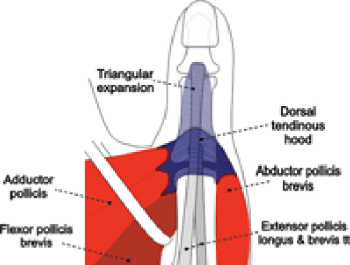 Mri Of Finger Tendons Radiology Key
 I Wear A Brace But It Still Hurts Part 3 The Elbow
 Pdf Cme Extensor Tendon Anatomy Injury And
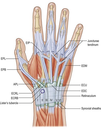 Extensor Tendon Injuries Plastic Surgery Key
 Extensor Tendon Injuries Plastic Surgery Key
 Injuries To The Hand And Digits Tintinalli S Emergency
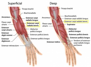 Lateral Epicondylitis Physiopedia
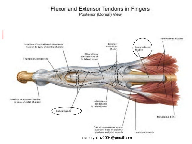 Extensor Tendons Injury And Deformity
 Extensor And Flexor Tendon Injuries In The Hand Wrist And
 Tendon Transfer And Grafting For Traumatic Extensor Tendon
 Extensor Tendon Injuries Florida Bone And Joint
 Finger Extensor Tendon Injury Rehab My Patient
 Extensor Tendon Injuries Pip Elson S Test Closing The Gap
 Extensor Tendons Of Pig Foot At Athabasca University Studyblue
 Extensor Tendon Lacerations Background History Of The
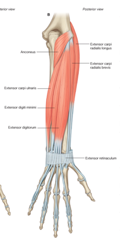
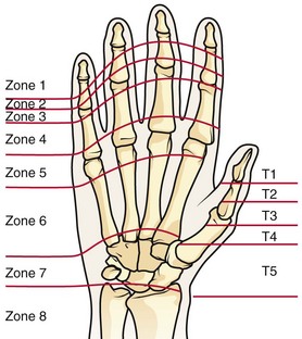 Extensor And Flexor Tendon Injuries In The Hand Wrist And
Patient Education Concord Orthopaedics
 The Muscles And Fasciae Of The Hand Human Anatomy
 Extensor Tendon Injuries Hand Orthobullets
 Metacarpophalangeal Joint Synovectomy And Extensor Tendon
Extensor Tendon Injuries Of The Finger Radsource
 Extensor Digitorum Muscle Wikipedia
 Extensor Tendon Repair Occupational Therapy Schools Hand
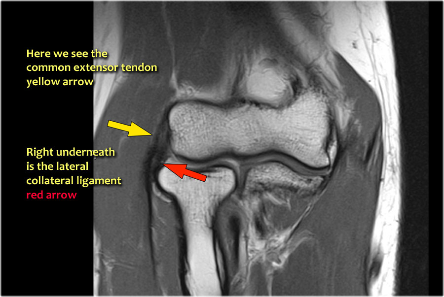 The Radiology Assistant Elbow Mri
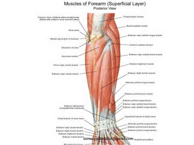 Extensor Tendons Injury And Deformity
Mri Of The Extensor Tendons Of The Wrist
Belum ada Komentar untuk "Extensor Tendon Anatomy"
Posting Komentar