Thoracic Cavity Anatomy
The thoracic wall actually encloses a cavity or space that is filled with various anatomical structures. Main lymphatic duct conveys blood to all lymph of lower limbs pelvic cavity abdominal cavity left thorax left arm and left side of head and neck thoracic duct crosses midline behind esophagus runs along left edge of esophagus enters root of neck turns inferiorly to cross the subclavian artery and enters the left brachiocephalic vein.
 Ch01 Body Cavity And Imaging Terms
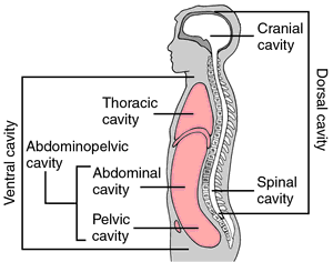
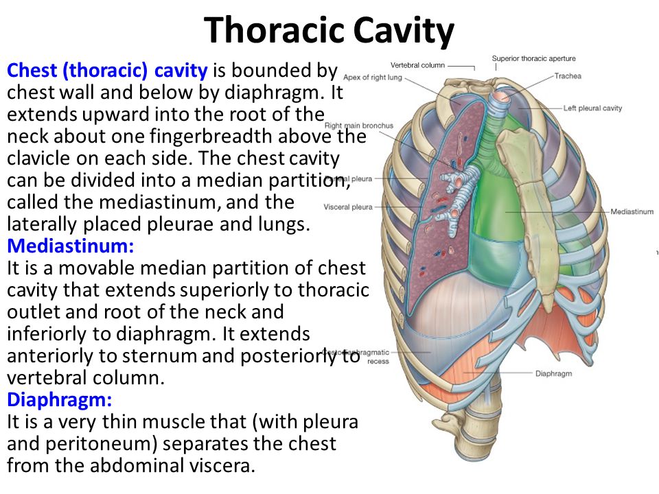
Divided by the diaphragm into the thoracic cavity and abdominopelvic cavity thoracic cavity.

Thoracic cavity anatomy. Contains the spinal cord which is an extension of the brain ventral cavity. Structures within the thoracic cavity include. The skeleton of the thorax and the shape and boundaries of the cavity.
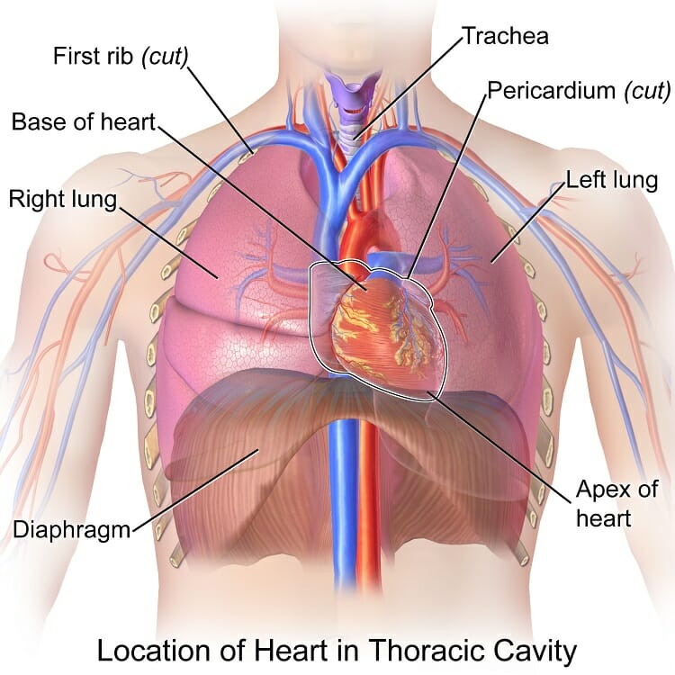
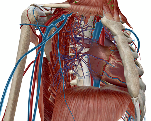
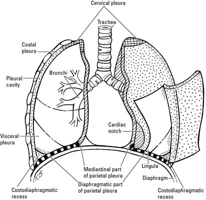
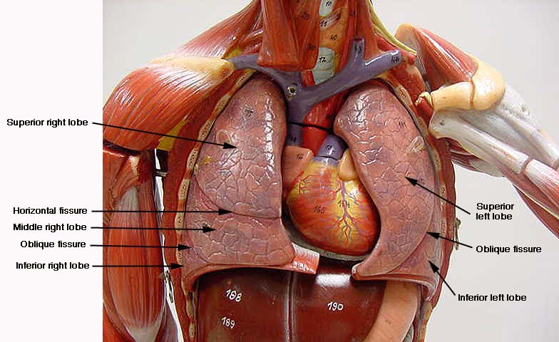
The heart lies between the two lungs and is enclosed within a fibrous bag the pericardium while each lung is invested by a serous membrane the pleura. Anatomy and physiology for dummies 3rd edition. Structures of the cardiovascular system including the heart and great vessels.
Structures of the respiratory system including the diaphragm trachea bronchi and lungs. The thoracic cavity the heart and lungs are situated in the thorax the walls of which afford them protection. Related biology terms abdominal cavity the cavity that lies posterior to the thoracic cavity and hold much of the digestive system.
Much of the focus when you explore the thoracic region is on the heart and lungs but the following organs also live in the thoracic cavity. The diaphragm is a dome shaped muscle that divides the thorax from the abdomen. Thoracic cavity also called chest cavity the second largest hollow space of the body.
Structures of the digestive system including the esophagus endocrine. Anterior portion of the torso. It is enclosed by the ribs the vertebral column and the sternum or breastbone and is separated from the abdominal cavity the bodys largest hollow space by a muscular and membranous partition the diaphragm.
Coelom the cavity or cavities formed during development that eventually become the thoracic and abdominal cavities. The pink lobed thymus is located between the. Contains the trachea bronchi lungs esophagus.
Now that weve covered the boundaries lets add another layer of knowledge to the initial basic thorax definition.
 Anatomy Lab Veins Of Thoracic Cavity Diagram Quizlet
 Thoracic Cavity Definition Organs Of Chest Cavity
 Thoracic Cavity Definition Of Thoracic Cavity By Medical
Thoracic Cavity Ii Heart And Great Vessels Anatomy 691
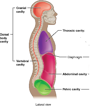 Dorsal Body Cavity Definition Organs Membranes Study Com
 Anatomy Of The Thoracic Wall Pulmonary Cavities And
 Surface Anatomy Of The Human Rv As It Lies In The Thoracic

 Anat20006 Lecture Notes Winter 2017 Lecture 24
 Anatomy And Physiology Anatomical Planes And Cavities
 Thoracic Wall And Breast Illustrations
 Opening The Thoracic Cavity
 Thoracic Cavity Contents Ppt Video Online Download
 What Is In The Thoracic Cavities Dummies

 Thoracic Cavity Mediastinum
 Chest Cavity
![]() Thorax Anatomy Wall Cavity Organs Neurovasculature
Thorax Anatomy Wall Cavity Organs Neurovasculature
 Anatomy Series The Thoracic Cavity By Dr Shakti Chandra
 Mediastinum Right Lateral View Right Thoracic Cavity And
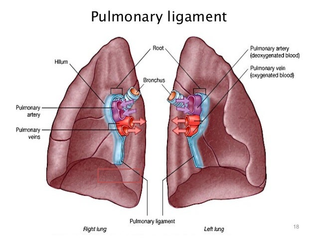
 Pulmonary Pleurae Wikipedia
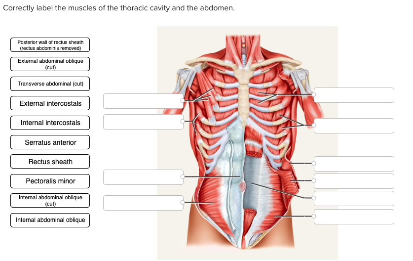 Solved Correctly Label The Muscles Of The Thoracic Cavity
 Anatomy Of The Thoracic Cavity 1st Plastic Model

 Anatomy Of The Chest Cavity Medical Art Works
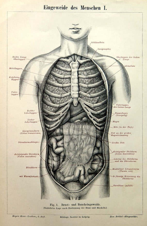 1894 Antique Organs Human Print Anatomy Body Engraving Vintage Viscera Old Lithograph Lungs Guts Thoracic Cavity Medical Wall Art
Belum ada Komentar untuk "Thoracic Cavity Anatomy"
Posting Komentar