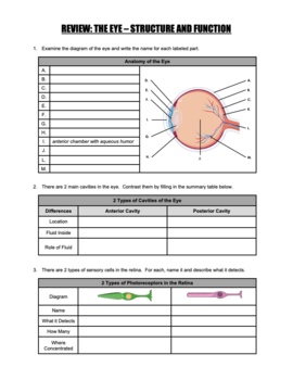
Anatomy Of The Eye And Functions
Behind the eye your optic nerve carries. And when there is low light the iris opens up the pupil to let in more light.
 Structure And Function Of The Eyes Eye Disorders Merck
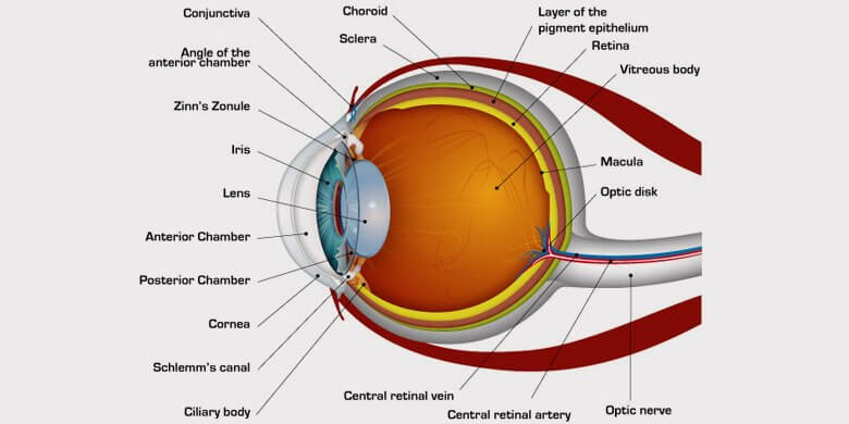
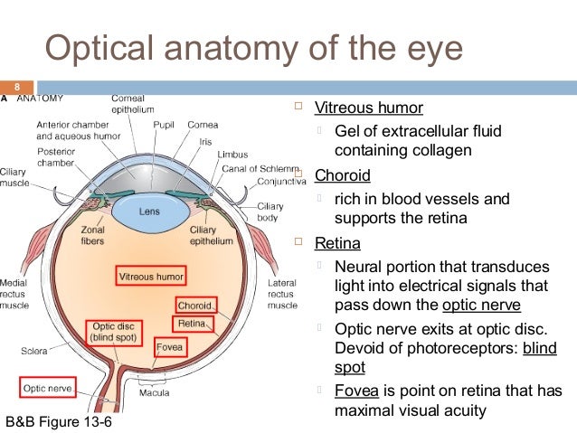
Optic disk connection of the optic nerve and retina also where the blood vessels enter eye.

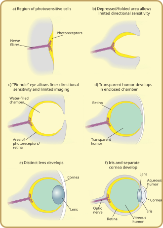
Anatomy of the eye and functions. It converts light into electrical impulses. Extraocular muscles help move the eye in different directions. But nevertheless apart from the obvious of eyesight and hearing they are responsible for various other activities.
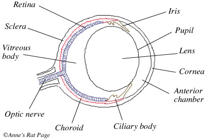
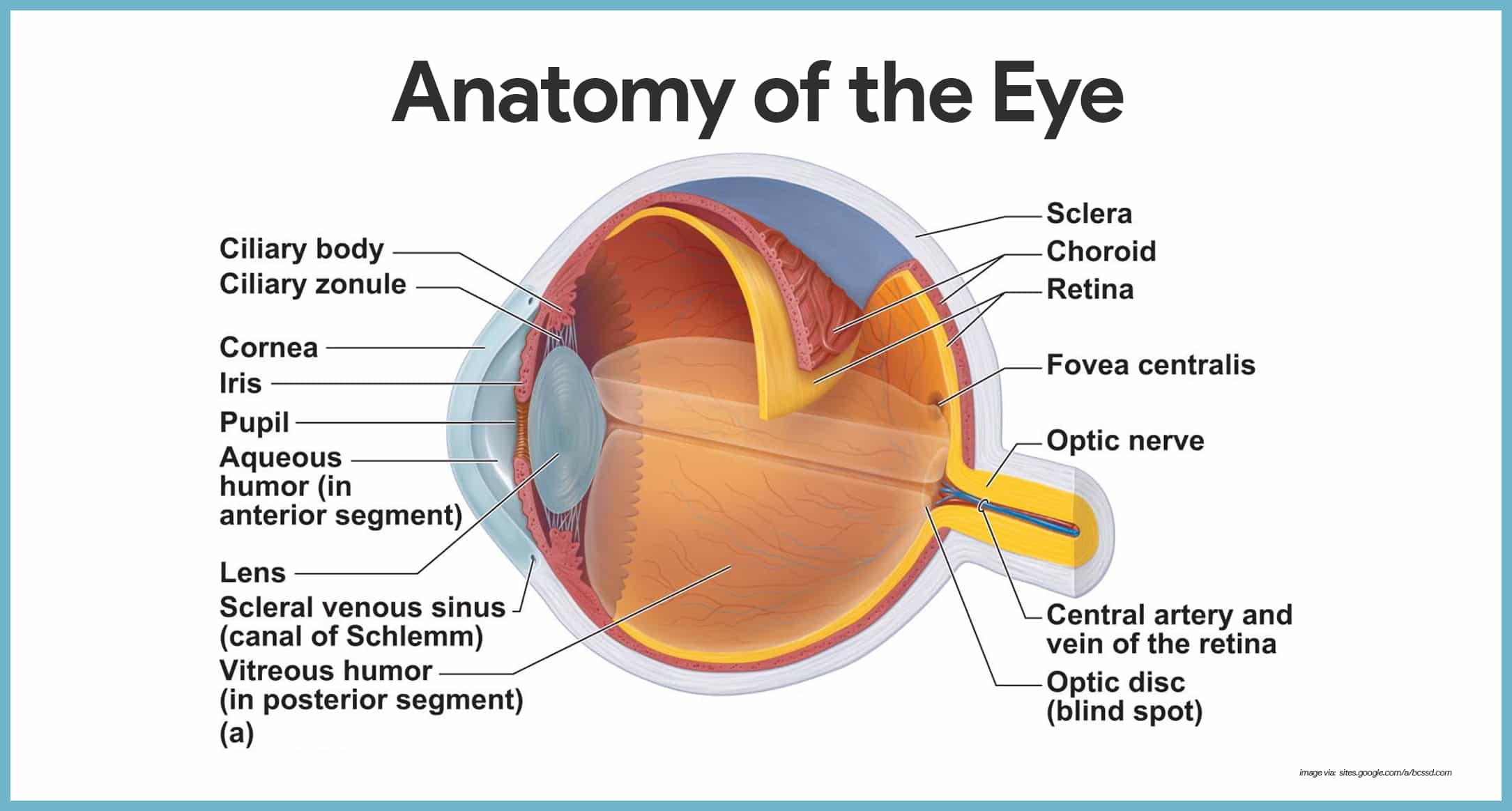
The eye has three main layers. Anatomy of the eye. Eye parts and functions.
These layers lie flat against each other and form the eyeball. In fact the ear is also responsible for maintaining your equilibrium or balance. The eye has many parts which work together to accomplish vision and to keep the structures required for vision safe from infection and injury.
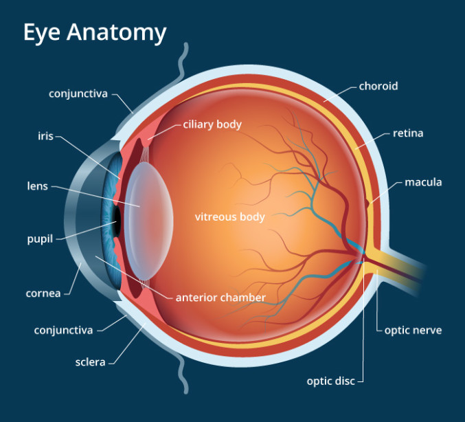
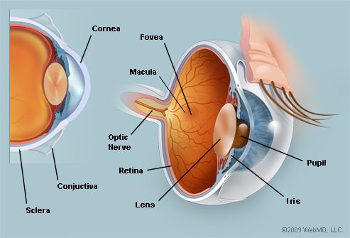
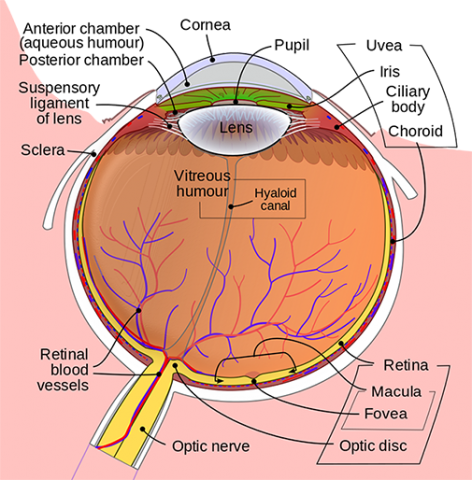
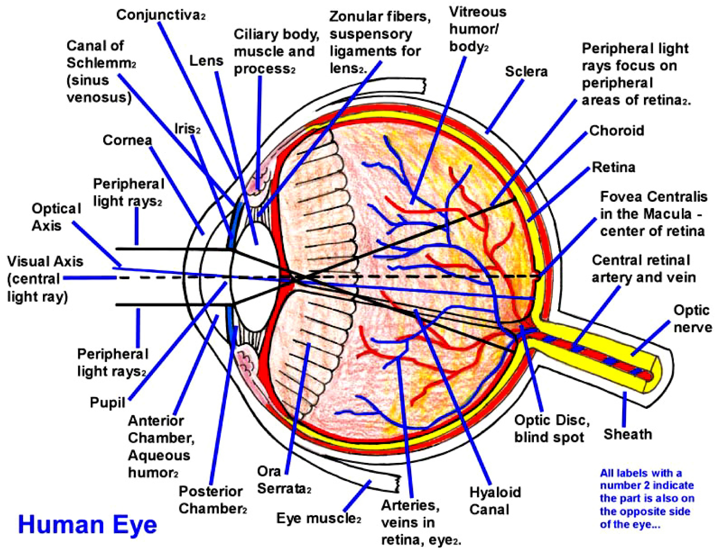
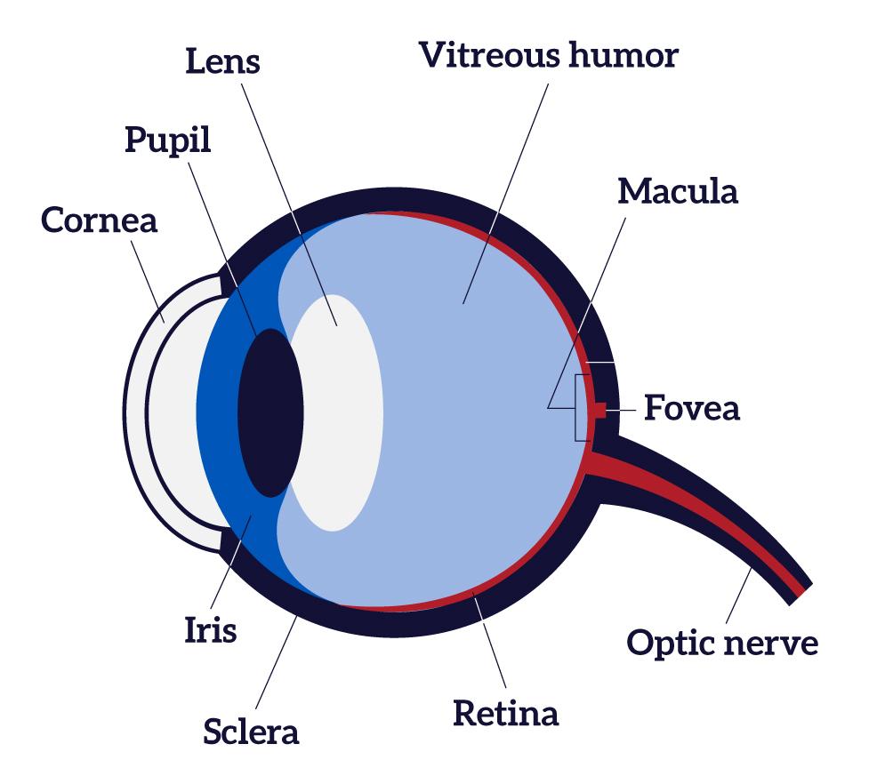
The pupil or black dot at the centre of the eye is an opening through which light can enter the eye. Nerve signals that contain visual information are transmitted through the optic nerve to the brain. The inside lining of the eye is covered by special light sensing cells that are collectively called the retina.
The outer layer of the eyeball is a tough white opaque membrane called the sclera the white of the eye. The sclera or white part of the eye protects the eyeball. The iris or coloured part of the eye surrounds the pupil.
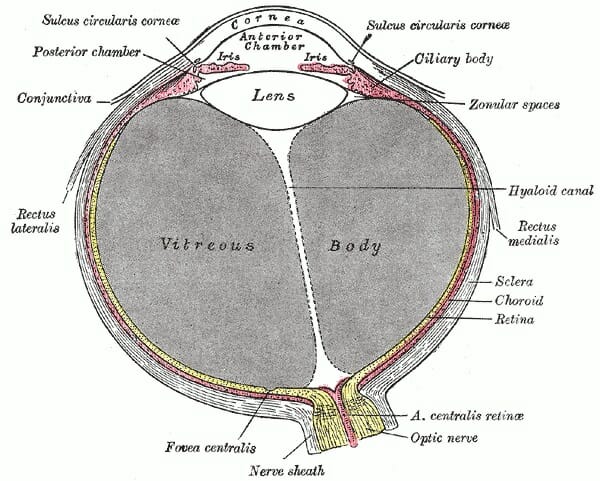
The eye is shaped like a round ball with a slight bulge at the front. The eye is surrounded by the orbital bones and is cushioned by pads of fat within the orbital socket. Anatomy of the eye.
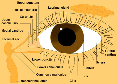
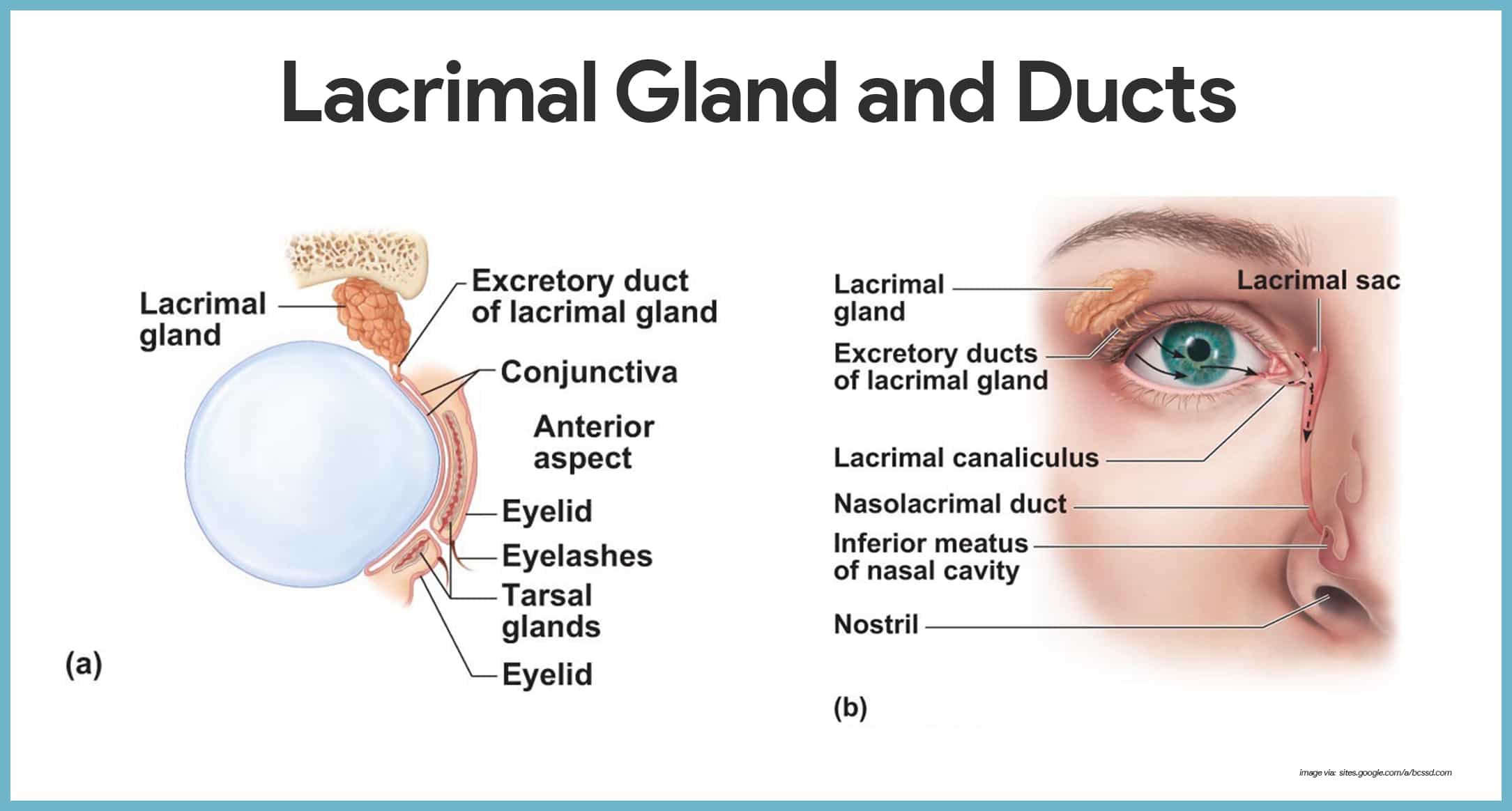
The surface of the eye and of the inner eyelids is covered by a clear protective membrane called the conjunctiva. Watery liquid inside the anterior chamber of the eye that helps maintain pressure and shape. To know more about the structure of eye and ear visit byjus.
Iris the colored part of the eye which helps regulate the amount of light entering the eye. When there is bright light the iris closes the pupil to let in less light. The anatomy of the eye.
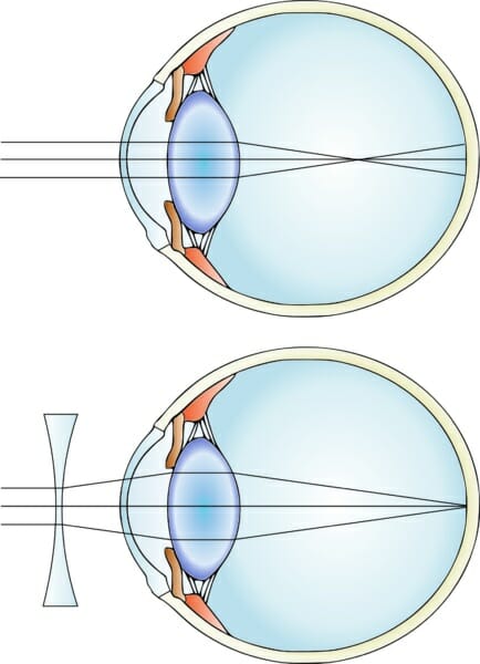
The functions of the eyes and ear need not be mentioned. The eye has many parts that must work together to produce clear vision. Lens focuses light rays onto the retina.
 Anatomy Of The Eye American Association For Pediatric
 Eye Wikipedia

 Eye Anatomy A Closer Look At The Parts Of The Eye
 Eyes Anatomy Overview Parts And Functions Biology
 How Do Your Eyes Work
 Eye Anatomy Structure Function Of Vision Hs Ls1 A
 Eye Anatomy Amazing Point Of View Eye Care
 Eye Structure And Function In Dogs Dog Owners Merck
 Anatomy Of The Eye Children S Wisconsin
 The Human Eye Facts Functions Structure And Problems
 Stuart R Winthrop M D Eye Anatomy And Function
 The Eyes Human Anatomy Diagram Optic Nerve Iris Cornea
 How Your Eyes Work
 Human Eye Ball Anatomy Physiology Diagram
 Anatomy Of The Eye Kellogg Eye Center Michigan Medicine
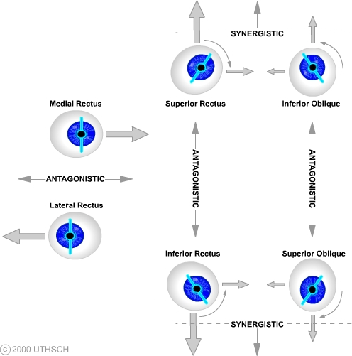 Ocular Motor Control Section 3 Chapter 8 Neuroscience
Eye Health Anatomy Of The Eye Visionaware
 Functions Of The Parts Of The Eye Eye Anatomy Parts Of
 Image Result For All The Parts Of The Eye And What They Do
 Human Eye 3d Poster Anatomy Wall Chart Updated Bump Design
 Anatomy Of The Eye Human Eye Anatomy Owlcation
Anatomy And Structure Of The Eye Brightfocus Foundation
 Special Senses Anatomy And Physiology Nurseslabs
 Special Senses Anatomy And Physiology Nurseslabs
 Eye Structure And Function In Dogs Dog Owners Merck
 How The Eyes Work National Eye Institute
 Eyes Anatomy Overview Parts And Functions Biology
 Visual System Structure And Function
Belum ada Komentar untuk "Anatomy Of The Eye And Functions"
Posting Komentar