Left Shoulder Anatomy
Mri of the shoulder. Orthopaedic surgical anatomy teaching collection add or remove collections home orthopaedic surgical anatomy teaching collection illustration of the left shoulder posterior view showing nerves arteries muscles and bones.
 Left Shoulder Normal Anatomy Medical Art Works
Anatomy of the left shoulder front view of normal shoulder anatomy with side view of joint opened.

Left shoulder anatomy. It stabilizes the shoulder and holds the head of the humerus in the glenoid a shallow cavity in the scapula. Download shoulder anatomy stock photos. This medical exhibit shows several images related to anatomy of the left shoulder.
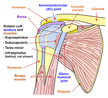
The collection of muscles and tendons in the shoulder is known as the rotator cuff. The shoulder joint is vulnerable to dislocations from sudden jerks of the arm especially in children before strong muscles have developed. Mr of the shoulder.
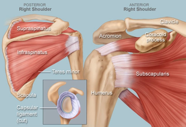
Inflammation of the bursa the small sac of fluid that rests over the rotator cuff tendons. Affordable and search from millions of royalty free images photos and vectors. The muscles of the rotator cuff include the suprasinatus infraspinatus teres minor and subscapularis.
Shoulder pain often occurs as a result of disease or injury that affects structures in the shoulder joint eg tendons ligaments muscles bones. Dislocation of the shoulder is extremely painful and may require surgical repair or even cause permanent damage. Muscles of the rotator cuff labeled on a sagittal mr slice.
Inflammation of one of the tendons in the shoulders rotator cuff. This medical image is titled anatomy of the left shoulder. Pain with overhead activities or pressure on the upper outer arm are symptoms.
Shoulder anatomy the shoulders are made up of bones cartilage muscles tendons and ligaments. An mri of the shoulder of a healthy subject was performed in the 3 planes of space coronal axial sagittal commonly used in osteoarticular imagery with two weightings most commonly used to explore.
 Shoulder Crane A Concept Of Suspension Stability Control
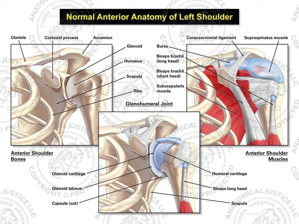 Anterior Anatomy Of The Left Shoulder
Left Shoulder Injuries High Impact Visual Litigation
 Latex Injected Left Shoulder Specimen Overview Of The Main
 Posterior View Of Left Shoulder Showing Paths Of Nerves From
 Shoulder Anatomy Shoulder Pain Info
 3d Printed Shoulder Left Superficial Muscles And Axillary Brachial Artery
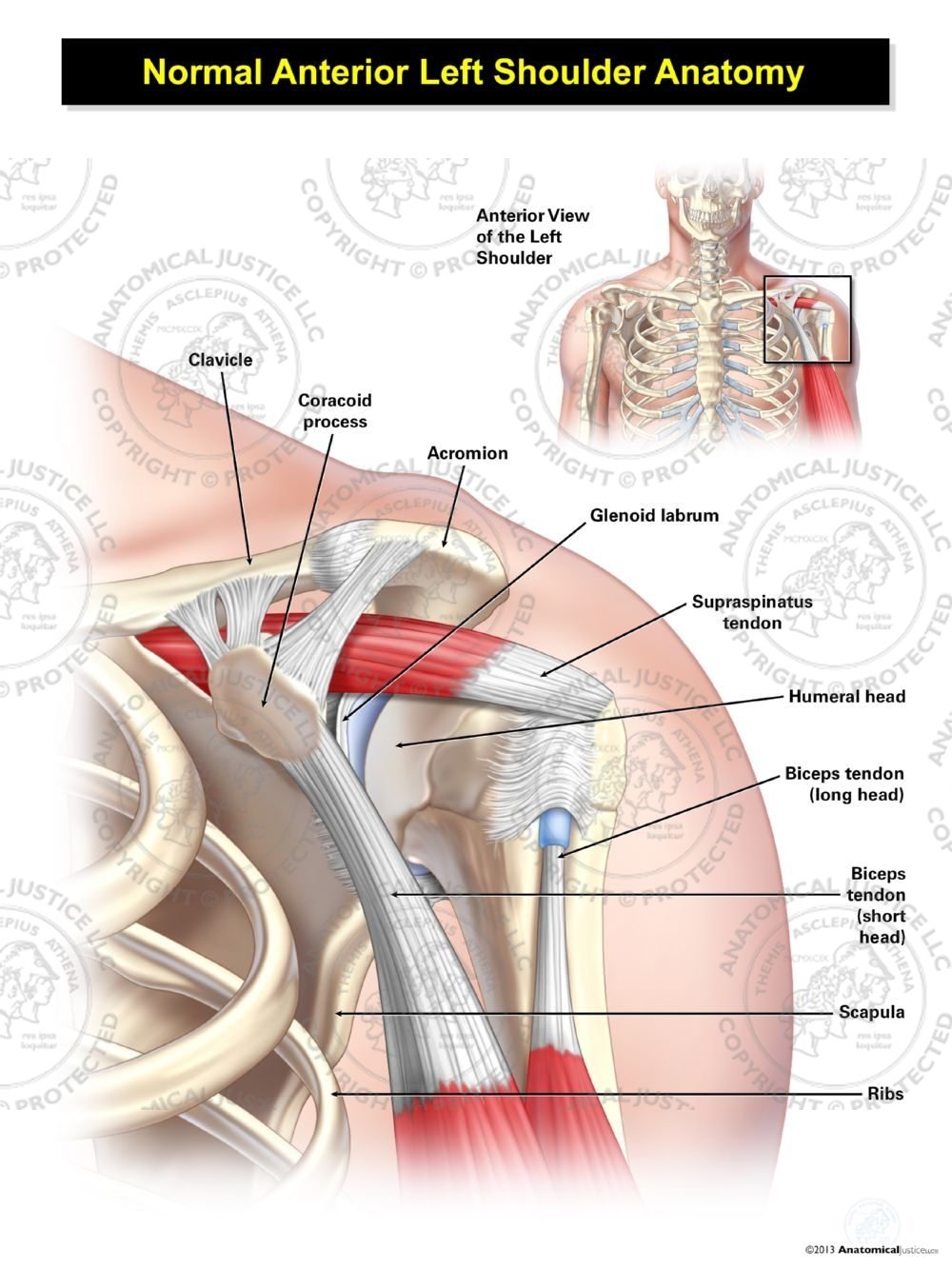 Normal Anterior Left Shoulder Anatomy

:max_bytes(150000):strip_icc()/shoulder-bones-and-muscles-971624580-9ac67b210b194ca6b414ffc28c8d3402.jpg) Anatomy Of The Human Shoulder Joint
Anatomy Of The Human Shoulder Joint
 Muscles Of The Shoulder Joint And Girdle Human Anatomy Kenhub
Frozen Shoulder Adhesive Capsulitis Orthoinfo Aaos
 Traumatic Injury To The Shoulder With Capsulitis Doctor Stock
 Shoulder Wikipedia
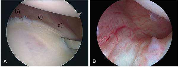
 Left Shoulder Injuries With Arthroscopic And Open Repairs
 Left Shoulder Injuries Pre Operative Condition Medical
 Shoulder Human Anatomy Image Function Parts And More
 Normal Left Shoulder Anatomy Of Muscles And Ligaments
![]() Anatomy Of Left Shoulder Coronal View Art Print
Anatomy Of Left Shoulder Coronal View Art Print
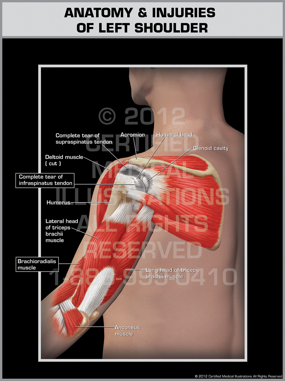 Anatomy Injuries Of Left Shoulder
 Anatomy And Biomechanics Of The Unstable Shoulder

![]() Fracture At Neck Of Humerus Arm Bone Film X Ray Left
Fracture At Neck Of Humerus Arm Bone Film X Ray Left
Search Left Shoulder Anatomy
Stock Image Illustration Of The Shoulder And The Rotator
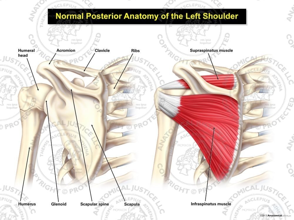 Normal Posterior Anatomy Of The Left Shoulder
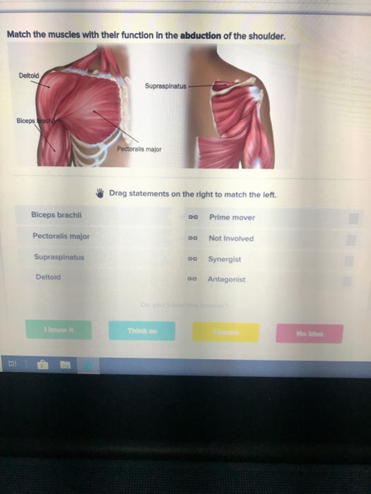 Solved Match The Muscles With Their Function In The Abduc
 Anatomy Moment The Shoulder Girdle Corpo Kinetic Pilates
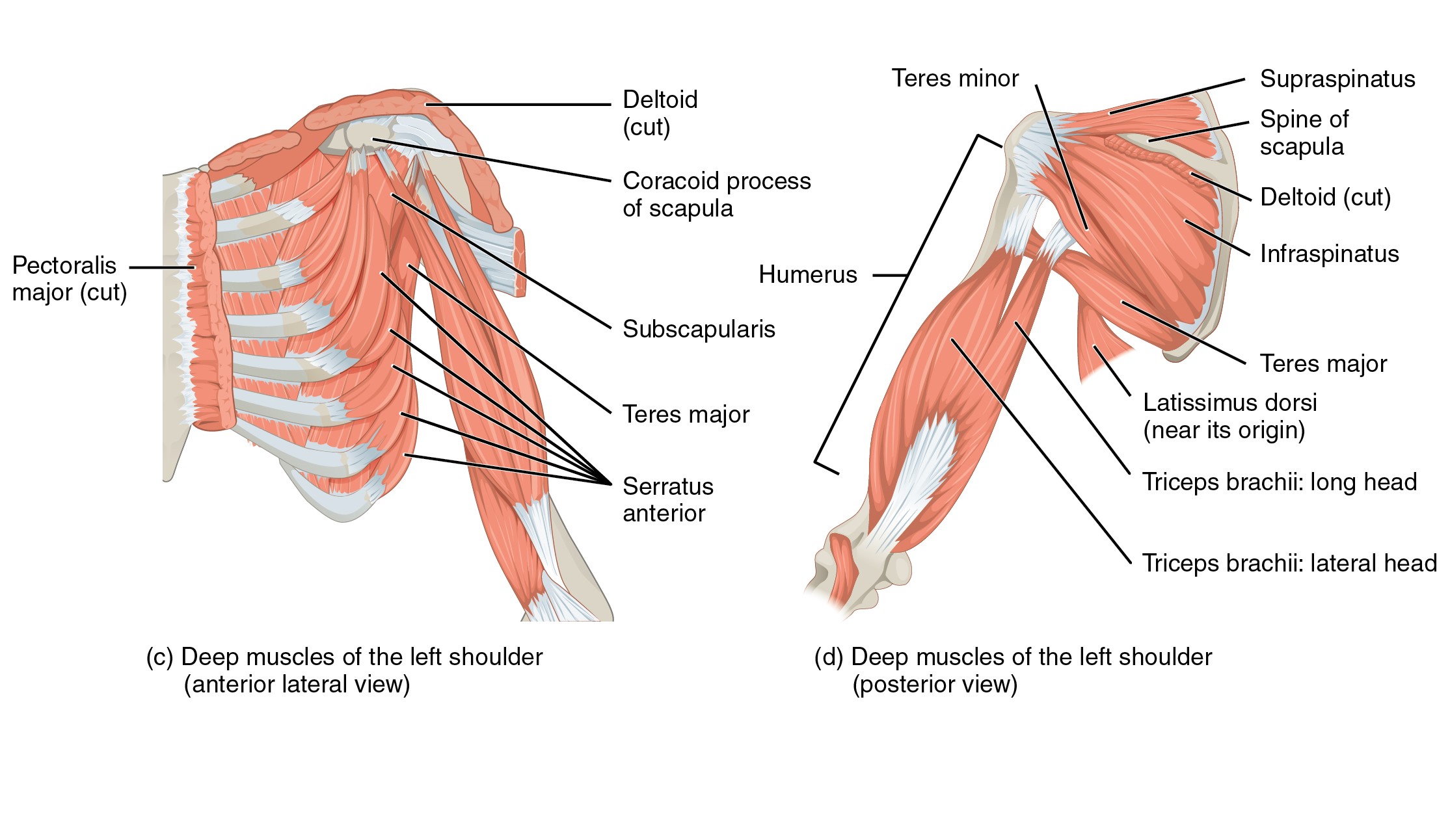
 Muscles Of The Pectoral Girdle And Upper Limbs Anatomy And
 Anatomy Of Left Shoulder Coronal View D1243 6 016

 This Trial Exhibit Shows The Rotator Cuff Muscles Of The
 Normal Left Shoulder Anatomy Anterior View Medical Art Works
Belum ada Komentar untuk "Left Shoulder Anatomy"
Posting Komentar