Oropharynx Anatomy
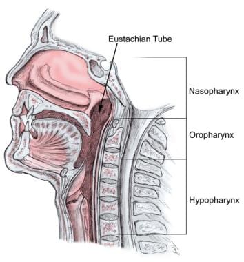
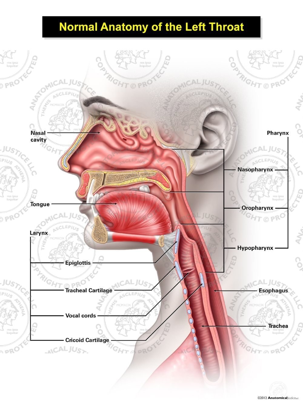
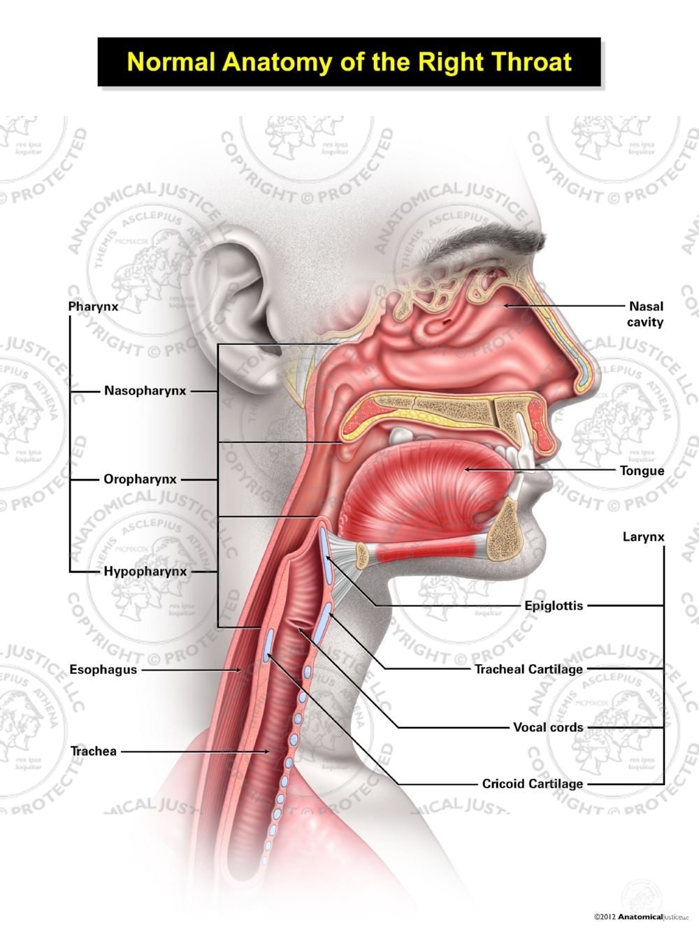
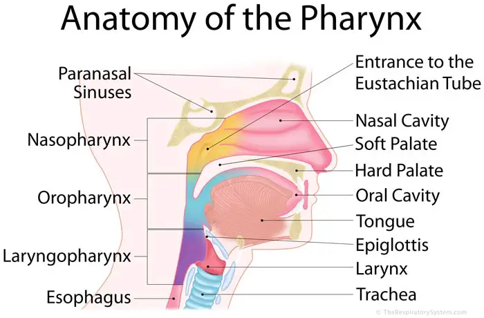
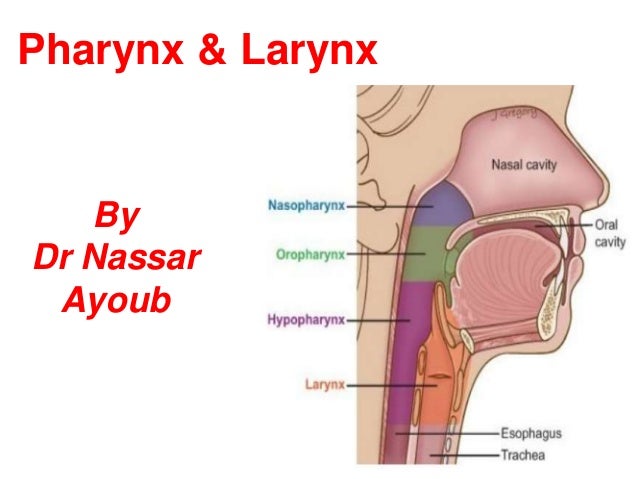
The oropharynx is behind the oral cavity below the soft palate and above the epiglottis. Anatomy of the oropharynx the pharynx is divided into the nasopharynx oropharynx and hypopharynx fig.
 Oropharynx Biopsy Overview Indications Contraindications
Oropharynx anatomy this shows the exact potion of the oropharynx just at the beginning of the esophagus showing.
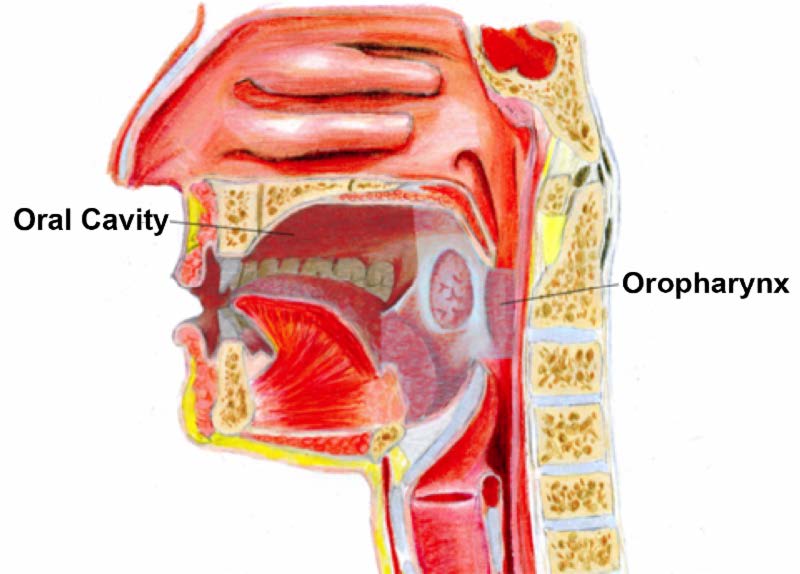
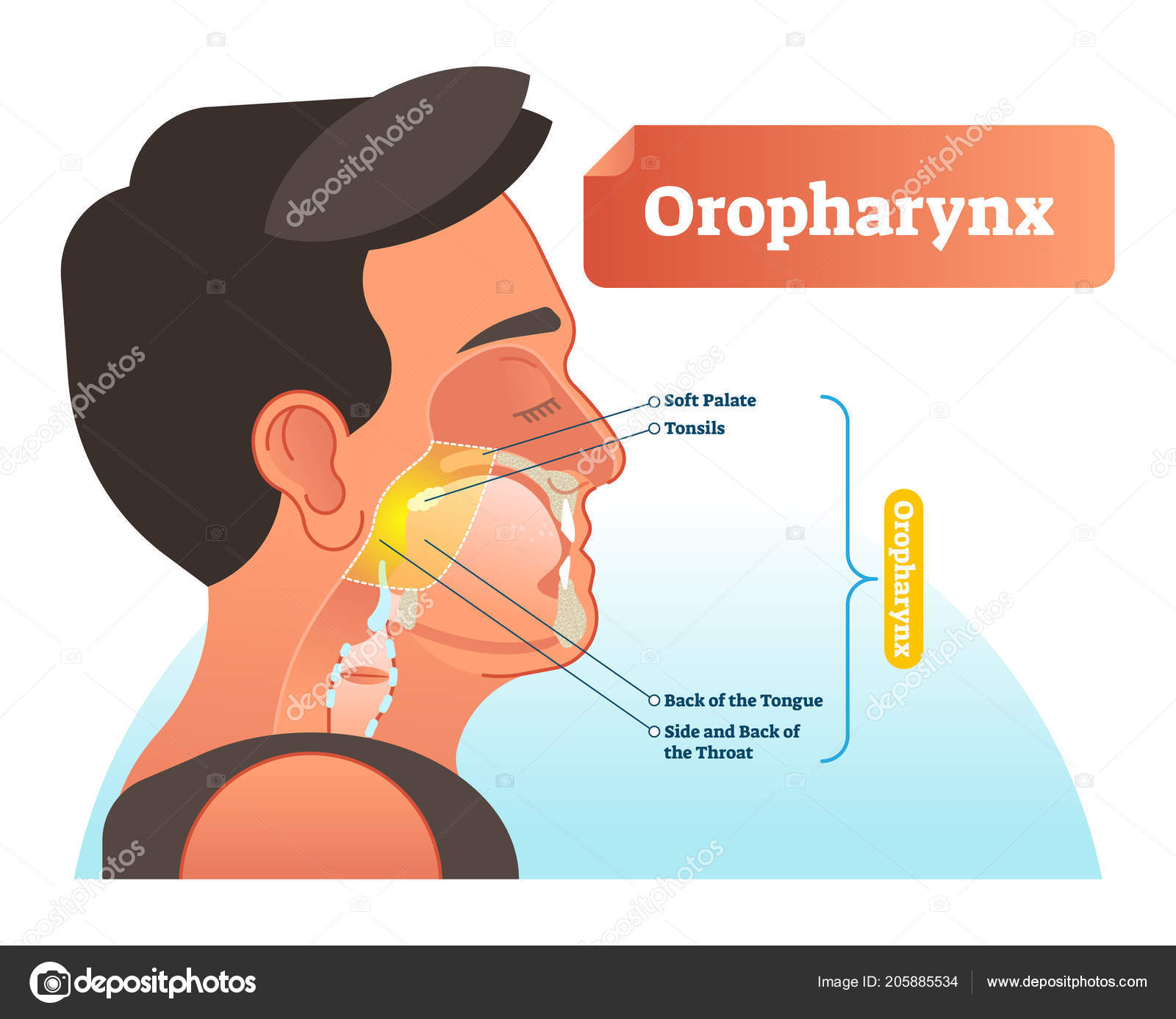
Oropharynx anatomy. The oropharynx includes the base of the tongue back 13 the soft palate at the back of the roof of the mouth the tonsils and the back of the throat. Anatomy the oropharynx is the posterior continuation of the oral cavity. Inserts posteriorly into to the pharyngeal tubercle of the occiput and the median pharyngeal raphe.
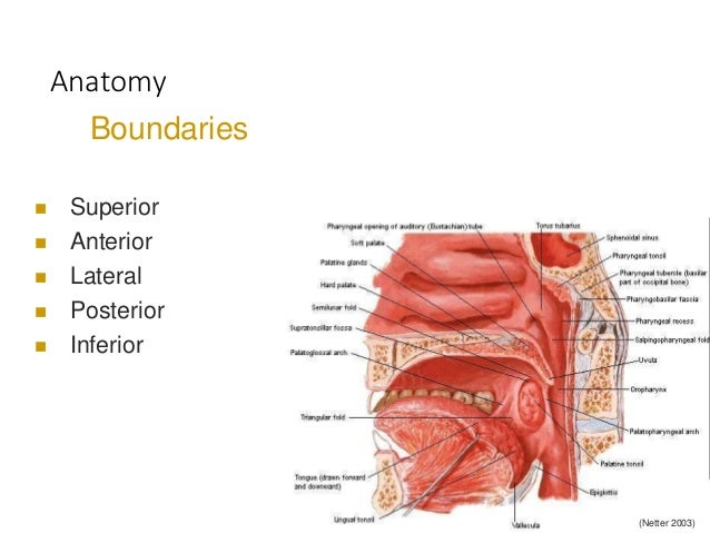
The superior wall consists of the inferior surface of the soft palate and the uvula. The oropharynx is the region posterior to the oral cavity that includes the posterior one third of the tongue tongue base the palatine tonsils the soft palate and the oropharyngeal mucosa and constrictor muscles. The posterior pharyngeal wall is at the level of the second and third cervical vertebrae.
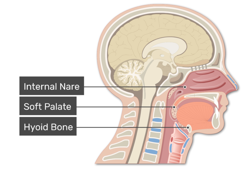
The oropharynx is located between the soft palate superiorly and the hyoid bone inferiorly. The lateral wall is made up of the tonsil tonsillar fossa and tonsillar faucial pillars. It communicates with the nasopharynx superiorly and the laryngopharynx inferiorly.
It is divided by the glossopalatine arch from the laryngeal pharynx. Because both food and air pass through the pharynx. It is continuous with the oral cavity anteriorly and communicates with the nasopharynx above and the supraglottic larynx and hypopharynx below.
The oropharynx forms part of the pharynx being the continuation of the oral cavity and nasopharynx superiorly and the larynx and hypopharynx inferiorly. Originates from the pterygomandibular ligament alveolar process of mandible and medial pterygoid plate and pterygoid hamulus of the sphenoid bone. Gross anatomy boundaries anteriorly.
The oropharynx is the middle section of the pharynx throat behind the mouth. The oropharynx has a stratified squamous epithelium. The lamina propria contains aggregates of lymphoid tissue called tonsils in several parts of the oropharynx.
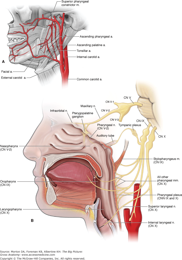
The oropharynx is innervated by the glossopharyngeal nerve. It is located in the oropharynx. It can be subdivided into the palatine faucial arch and oropharynx proper.
The oropharynx is the middle part of the pharynx directly below the soft palate that communicates anteriorly with the oral cavity proper by the isthmus of the fauces also known as the oropharyngeal isthmus. Vertical plane defined by the circumvallate papill. It is keratinised in ruminants.
The anterior wall consists of the base of the tongue and the epiglottic vallecula.
 Anatomical Regions Of The Pharynx
 Staging System For Hpv Throat Cancer Head And Neck Cancer
 Pharynx Wikipedia
 Oropharynx Stock Photos Oropharynx Stock Images Alamy
 Oropharynx An Overview Sciencedirect Topics
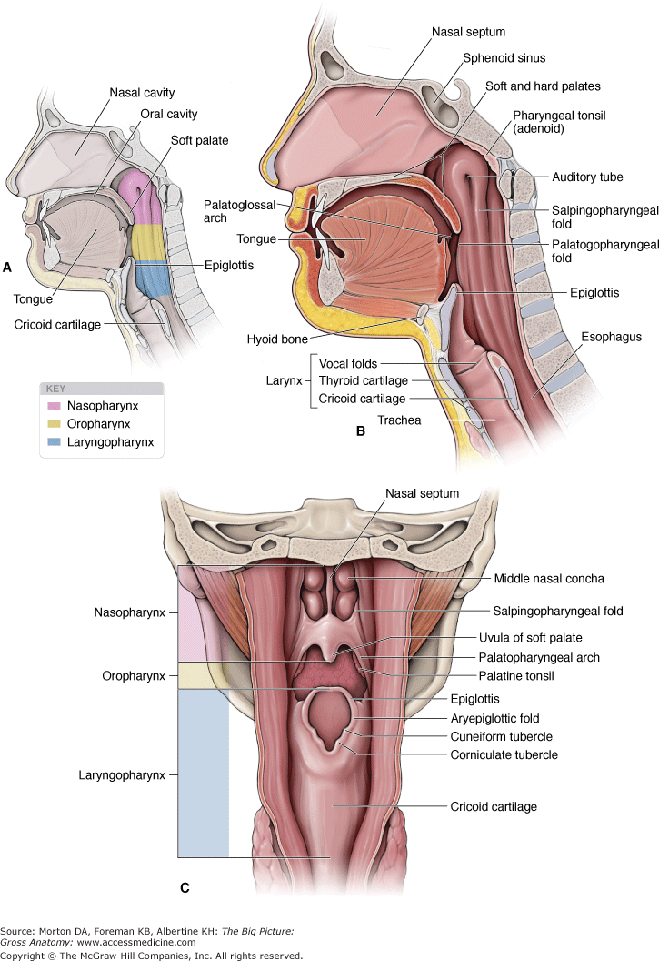 Chapter 27 Pharynx The Big Picture Gross Anatomy
 Oropharynx Images Stock Photos Vectors Shutterstock
 Tumors Of The Oropharynx Ento Key
Chapter 27 Pharynx The Big Picture Gross Anatomy
 Anatomy Of Tonsil Cancer Headandneckcancerguide Org
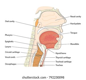
 Pharynx Larynx Anatomical Chart
 What Is Pharynx 25 Amazing Facts About Pharynx Parts
 Ca Oropharynx
![]() Pharynx Throat Anatomy Muscles Arteries And Nerves Kenhub
Pharynx Throat Anatomy Muscles Arteries And Nerves Kenhub
 Pharynx Anatomy Britannica
 Throat Diagram Labeled Oropharynx Vector Illustration

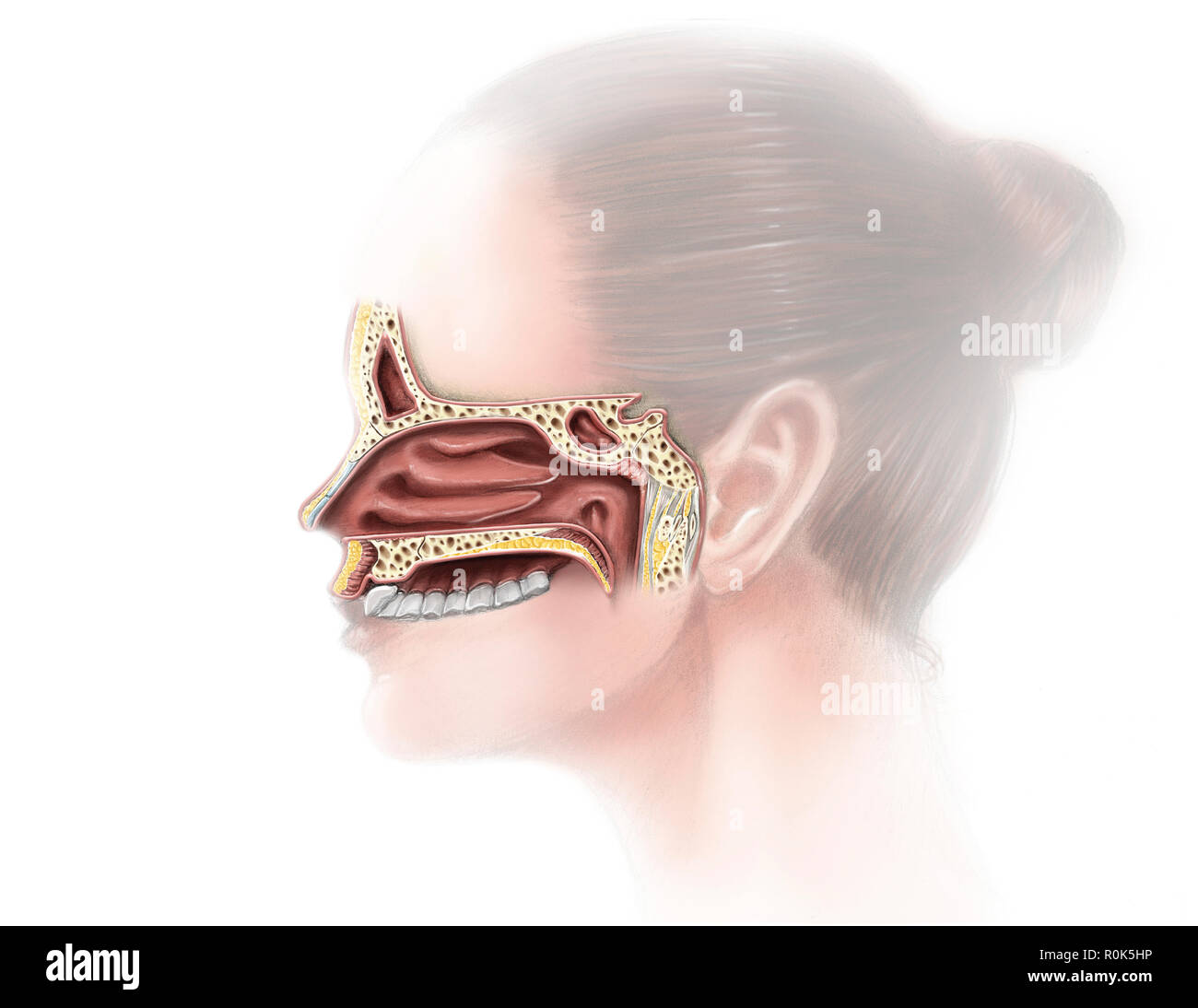
 Normal Female Anatomy Of The Left Throat
 Normal Male Anatomy Of The Right Throat
 Oral Cavity And Oropharynx
5 1 Pharynx Lecture Note 13 Hlth213 Mq Studocu
 Where Are The Larynx Pharynx And Trachea Located Which Of
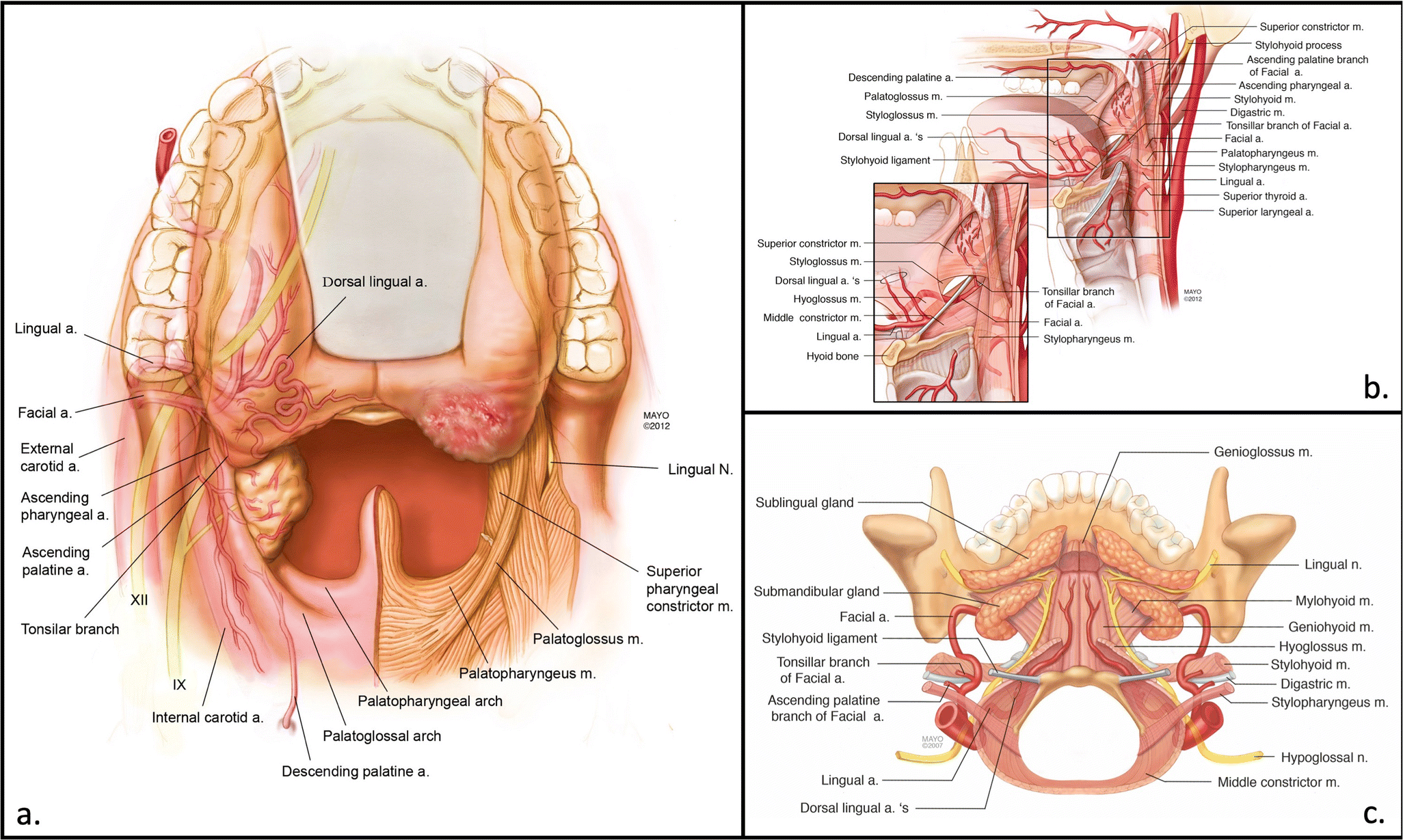
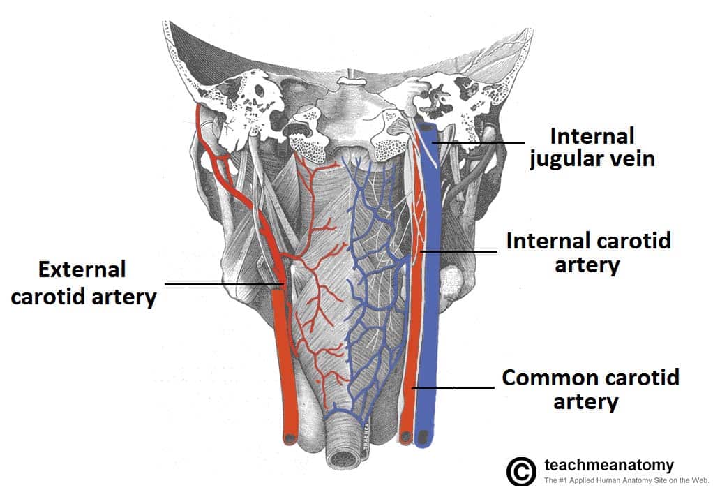 The Pharynx Subdivisions Blood Supply Teachmeanatomy
 Pharynx Definition Anatomy Functions And Diagram

 Pharynx Pharynx Prof Saeed Makarem Ppt Video Online Download
 Anatomy Of Pharynx Larynx
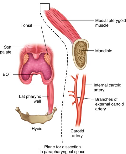
 Transoral Robotic Surgery Tors Inside Out Anatomy And
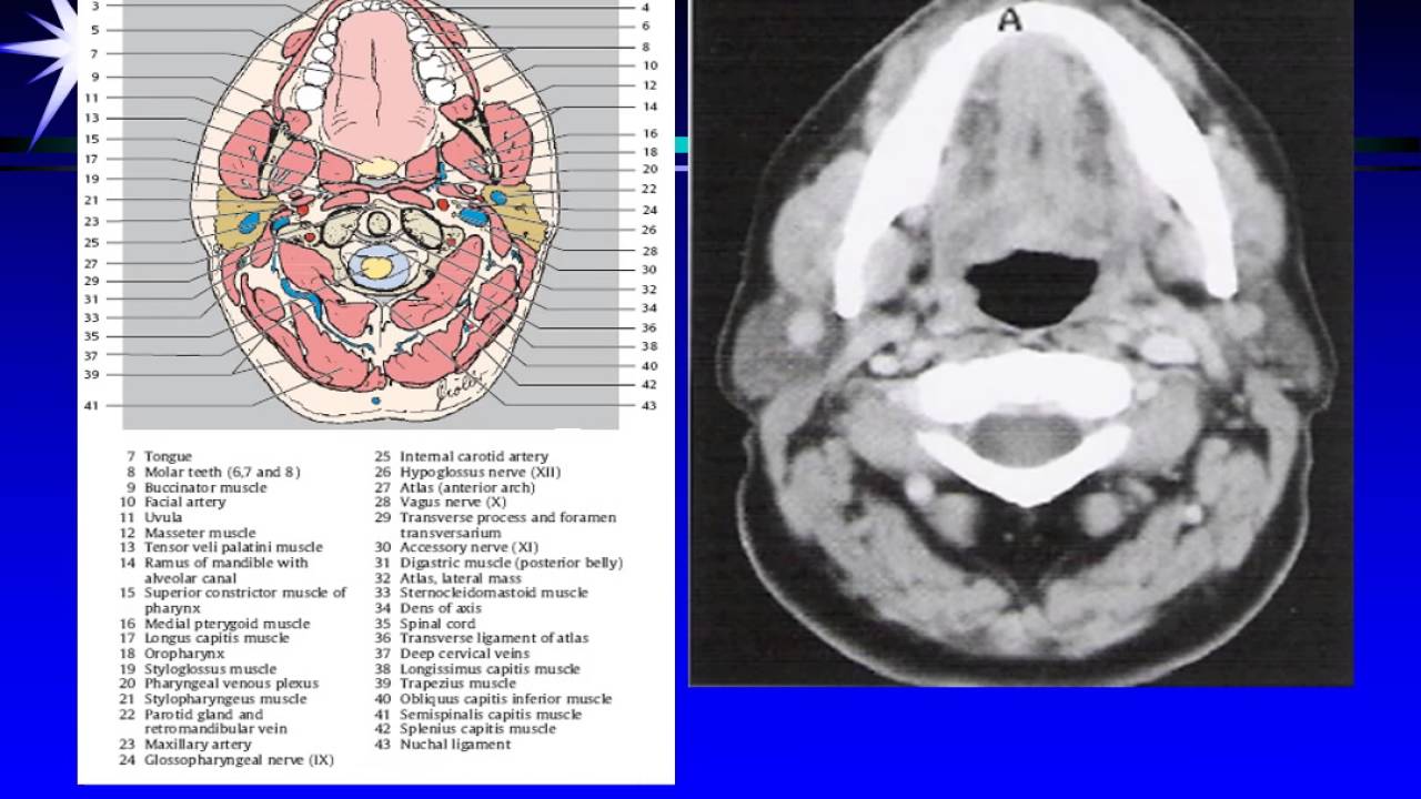 Ct Mri Larynx Pharynx Anatomy
Belum ada Komentar untuk "Oropharynx Anatomy"
Posting Komentar