Rectal Anatomy
This is commonly caused by a weakened pelvic floor after childbirth. Anatomy the rectum is a hollow muscular tube about 8 inches 20 cm in length and 25 inches in diameter at its widest point.
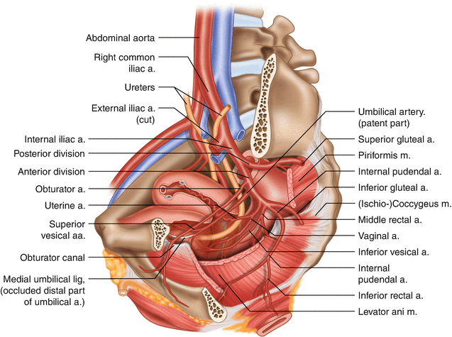 Rectal Anatomy Clinical Perspective Springerlink
An internal sphincter muscle which can be felt as a muscular ring beyond which is the rectum.

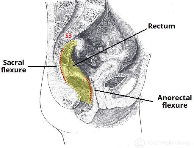
Rectal anatomy. The anus is the opening where the gastrointestinal tract ends and exits the body. The rectum is the most distal segment of the large intestine and has an important role as a temporary store of faeces. At the level of the s3 vertebral body the sigmoid colon loses its mesentery.
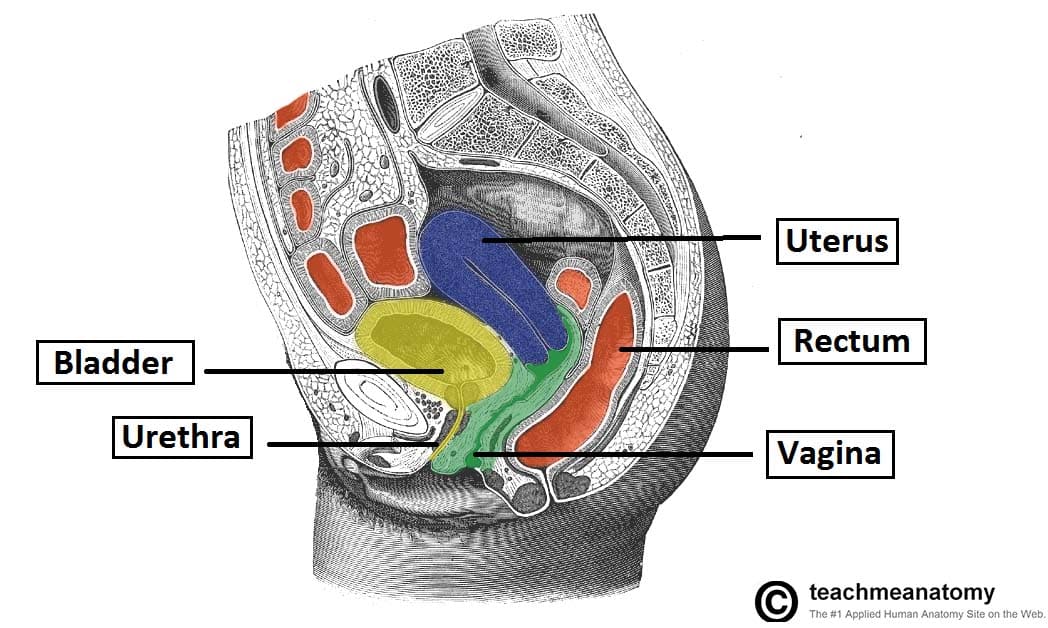
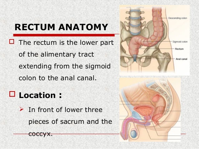
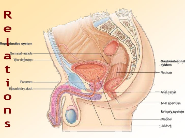
Rectal prolapse referring to the prolapse of the rectum into the anus or external area. It extends from the inferior end of the sigmoid colon along the anterior surface of the sacrum and coccyx in the posterior of the pelvic cavity. It is continuous proximally with the sigmoid colon and terminates into the anal canal.
The anus starts at the bottom of the rectum the last portion of the colon large intestine. A muscular sheet called the pelvic diaphragm runs perpendicular to the juncture of the rectum and rectum terminal segment of the digestive system in which feces accumulate just prior to discharge. The anorectal line separates the anus from the rectum.
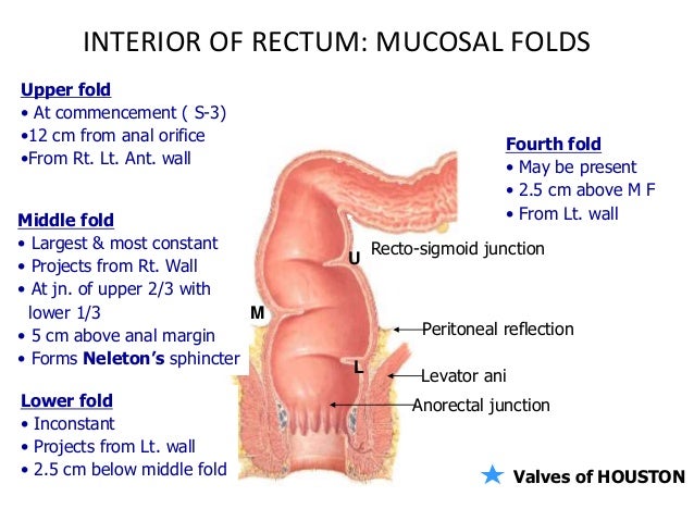
The columns are vascular and enlargement of their venous plexus results in internal hemorrhoids. The peritoneum firmly attaches the rectum to the sacrum. Anatomy of the anus.
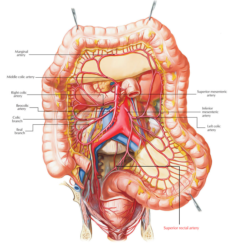
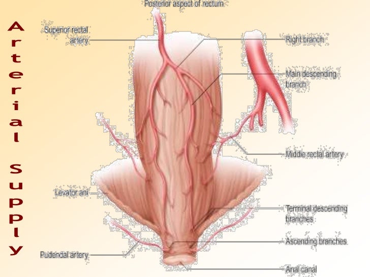
Body temperature may also be obtained from the rectal area. Veins corresponding to their named arteries form a rectal venous plexus. There are two sphincter muscles.
In this article we will discuss the anatomy of the rectum its structure anatomical relationships and clinical relevance. The rectum is an expandable organ for the temporary storage of feces. Several vertical mucosal folds the anal formerly called rectal columns are usually visible in the upper half of the canal fig.
Certain types of cancers may be diagnosed by performing an endoscopy in the rectum. Arterial supply to the rectum is formed from an anastomotic submucous plexus. An endoscopy is a procedure where a doctor uses an endoscope a small flexible tube with a camera and light to examine areas inside the body.
The anal columns are united below by anal valves which bound anal sinuses. Tough tissue called fascia surrounds the anus and attaches it to nearby structures. And an external sphincter muscle.
The upper portion of the anus or that part that connects to the rectum is known as the squamocolumnar junction. The rectum is continuous with the sigmoid colon and extends 13 to 15 cm 5 to 6 inches to the anus. The rectum lies next to the sacrum and generally follows its curvature.
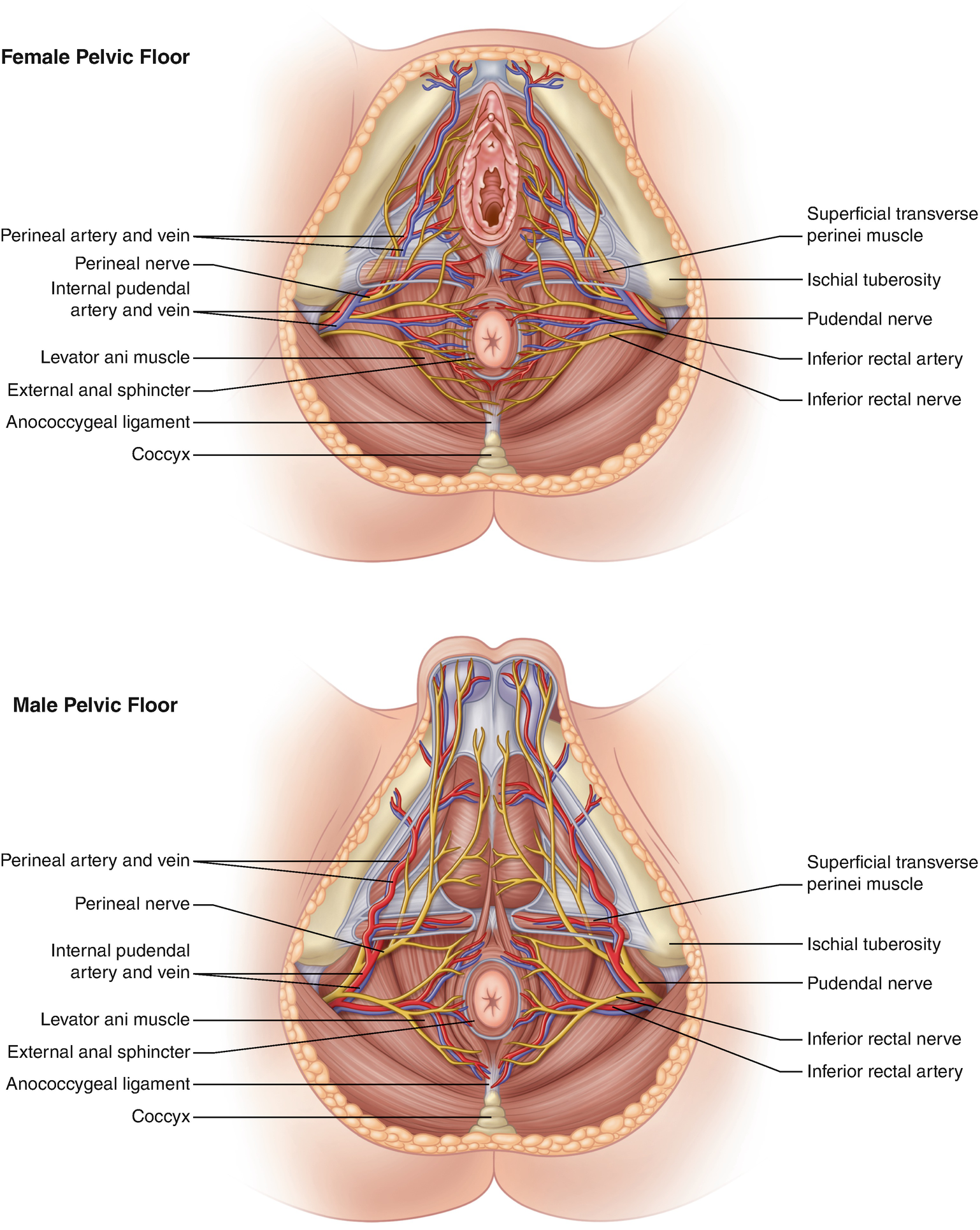 Anatomy And Embryology Of The Colon Rectum And Anus
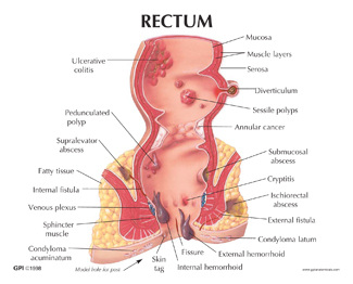 Function And Anatomy Of The Rectum Health With Nature
 Anal Canal An Overview Sciencedirect Topics
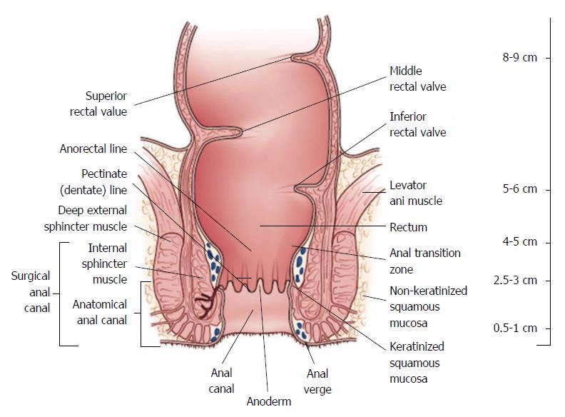 Rectal Cancer An Evidence Based Update For Primary Care
 The Rectum Position Neurovascular Supply Teachmeanatomy
 Superior Rectal Artery Wikipedia
:watermark(/images/watermark_only.png,0,0,0):watermark(/images/logo_url.png,-10,-10,0):format(jpeg)/images/anatomy_term/plicae-transversae-recti/0uznSNwBULsxINbpa1zbg_Transverse_folds_of_rectum_01.png) Rectum Anatomy Histology Function Kenhub
Rectum Anatomy Histology Function Kenhub
 Human Papilloma Virus And Squamous Cell Carcinoma Of The
 The Rectum Position Neurovascular Supply Teachmeanatomy
 Radiotherapy Dictionary Rectal Division
 Ch 20 Anus Rectum Prostate At University Of South
 Superior Rectal Artery Earth S Lab
 Anatomy Of The Pelvic Autonomic Nerves With Relation To
 Rectum Model Of The Human Anatomy Pathological Disease Rectal Ulcer Acne Pathological Model Use For Medical Anatomical Model
:background_color(FFFFFF):format(jpeg)/images/library/10928/the-rectum-and-anal-canal_english.jpg) Rectum Anatomy Histology Function Kenhub
Rectum Anatomy Histology Function Kenhub
 Hsf Anatomy Of Rectum And Anus Flashcards Quizlet
:watermark(/images/watermark_5000_10percent.png,0,0,0):watermark(/images/logo_url.png,-10,-10,0):format(jpeg)/images/atlas_overview_image/813/nMWLMYwVgSet78uvB3fng_the-blood-vessels-of-the-rectum_english.jpg) Rectum Anatomy Histology Function Kenhub
Rectum Anatomy Histology Function Kenhub
 Rectal Prolapse Expanded Version Ascrs
 Zgood Medical Anatomical Rectal Anal Canal Structure Mmodel
 Rectum And Anal Canal Anatomy And Function Preview
 Anatomy And Cell Biology 3319 Lecture Notes Fall 2017
 The Colon What It Is What It Does Ascrs
 Anal Canal An Overview Sciencedirect Topics

Belum ada Komentar untuk "Rectal Anatomy"
Posting Komentar