Foot Bottom Anatomy
The foot is made up of 26 bones 33 joints and over 100 ligaments tendon and muscle what you will learn in the article. Again thank you from the bottom of my heart for taking the time to answer my questionyour an angel.
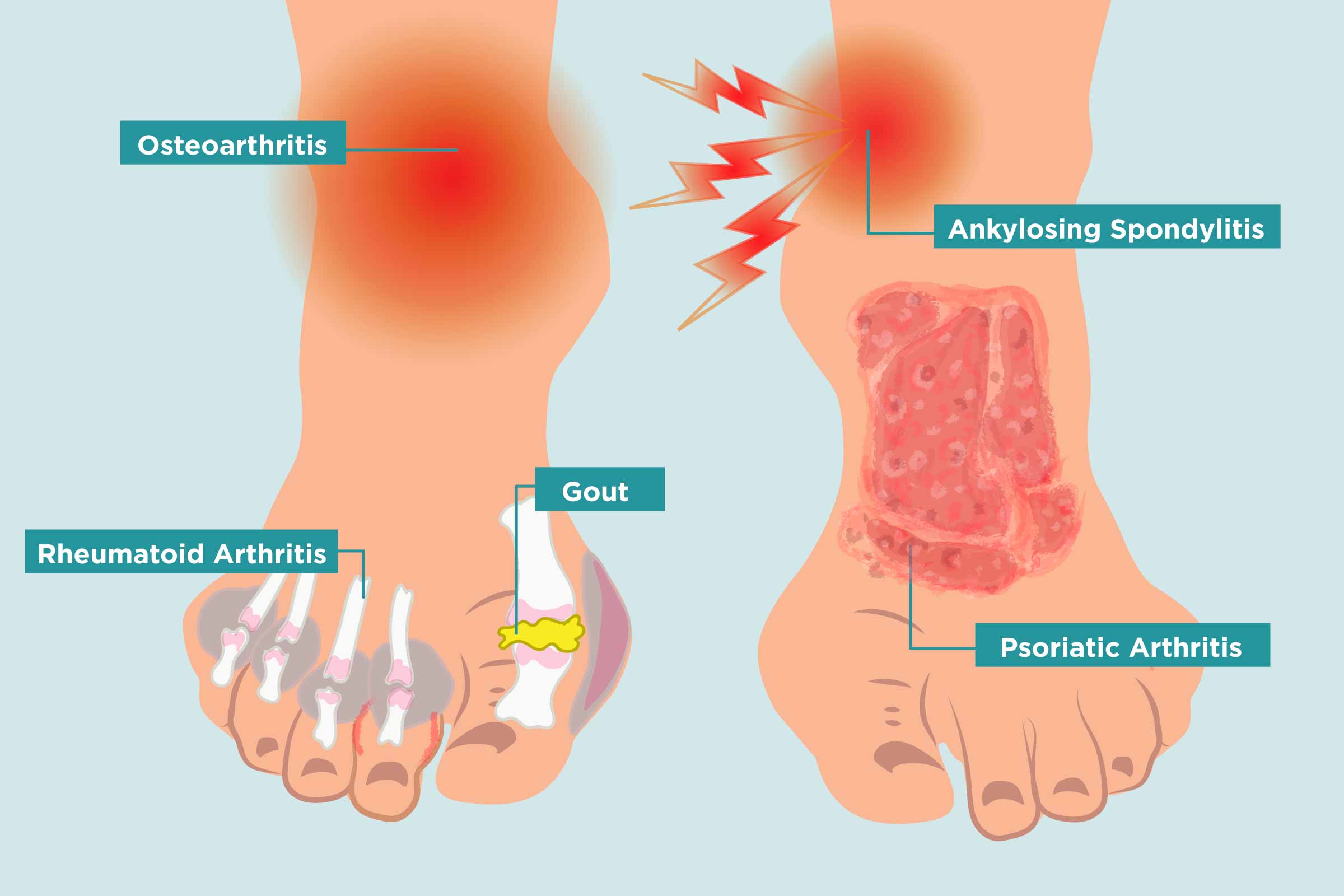 Arthritis In Your Feet Causes Symptoms And Treatment
Use it to pin point where you are having foot or ankle pain.
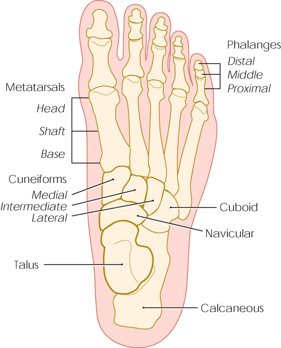
Foot bottom anatomy. The foot is an extremely complex anatomic structure made up of 26 bones and 33 joints that must work together with 19 muscles and 107 ligaments to execute highly precise movements. The skin on the bottom of our feet protects our muscles bones tendons and ligaments from injury. Therefore plantar refers to the bottom aspect of the foot.
It also prevents infection. Marc mitnick dpm. Each foot is made up of 28 bones 30 joints and more than 100 muscles tendons and ligaments all of which work together to provide support balance and mobility.
In humans the sole of the foot is anatomically referred to as the plantar aspect. These compressed patches of dead skin cells can be hard and painful. The feet are flexible structures of bones joints muscles and soft tissues that let us stand upright and perform activities like walking running and jumping.
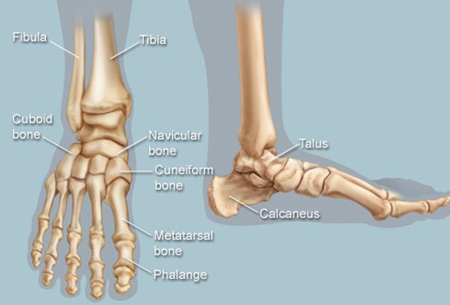
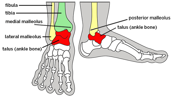
It slopes upward to meet the tarsal bones which point downward along with the remaining bones of the. Diagram of normal foot and ankle anatomy. Foot anatomy reference author.
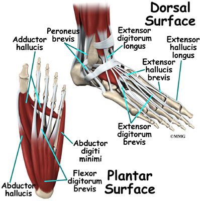
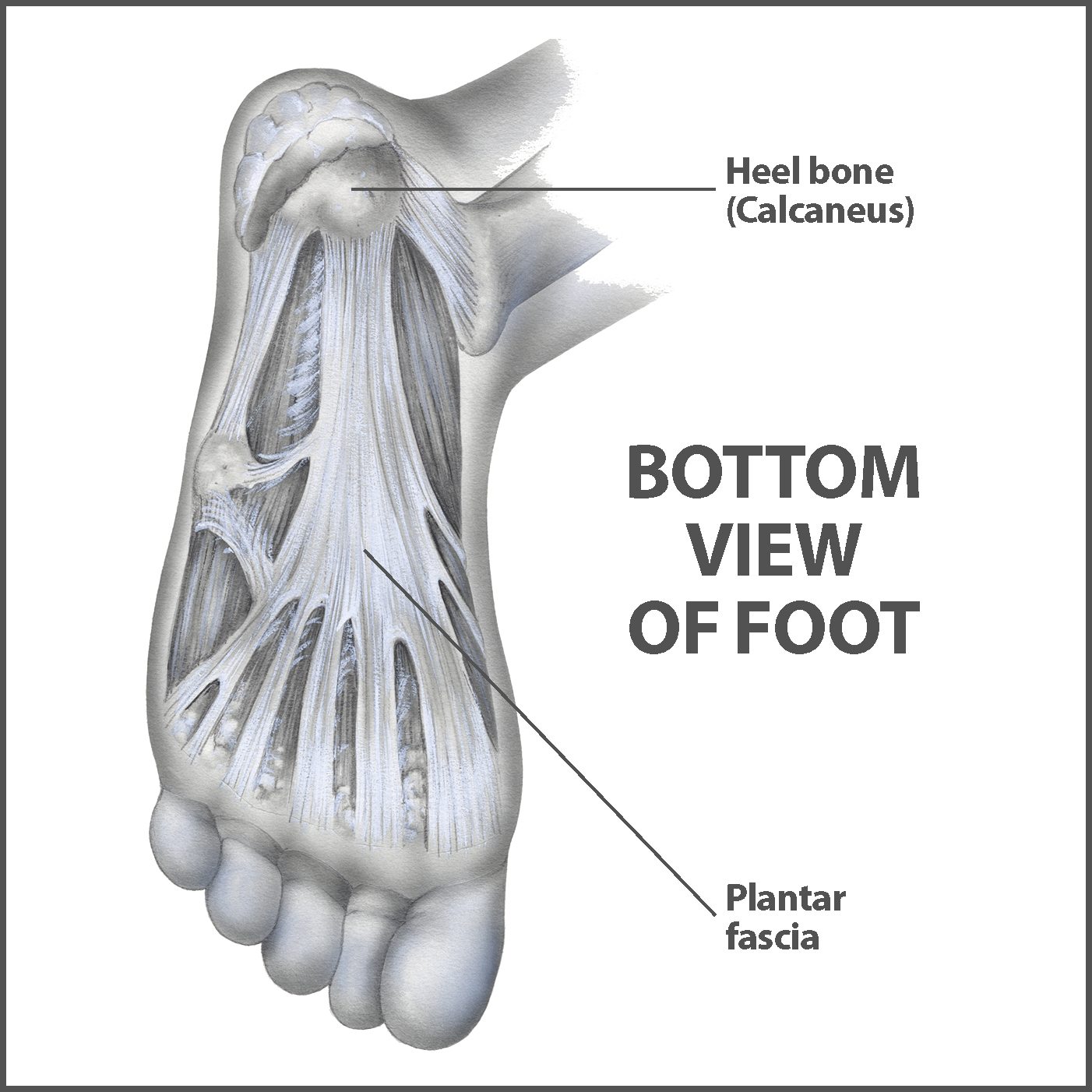
The feet are divided into three. The largest bone of the foot the calcaneus forms what is commonly referred to as the heel. Dorsal and plantar see fig.
The end of the leg on which a person normally stands and walks. The anatomy of the foot anatomi cally the foot has two surfaces. Also for students and health professionals it is critical to understand the foot anatomy which basically improves your knowledge of bone position ligament attachment and the way tendons run on the foot bones.
Anatomy of the foot an inside look at the structure of the foot. Calluses form on the bottom of the foot especially under the heels or balls and on the sides of toes. Dorsal refers to the top surface of the foot whereas plantar takes its name from the fact that the foot is planted on the ground when it is in contact with a surface.
The sole is the bottom of the foot. Toenails protect the top of our toes which as we all know can sometimes be vulnerable to being stubbed stepped on or having things dropped on them. Heres a look at the main structures of the feet.
 Nerve Blocks Of The Foot And Ankle Down East Emergency
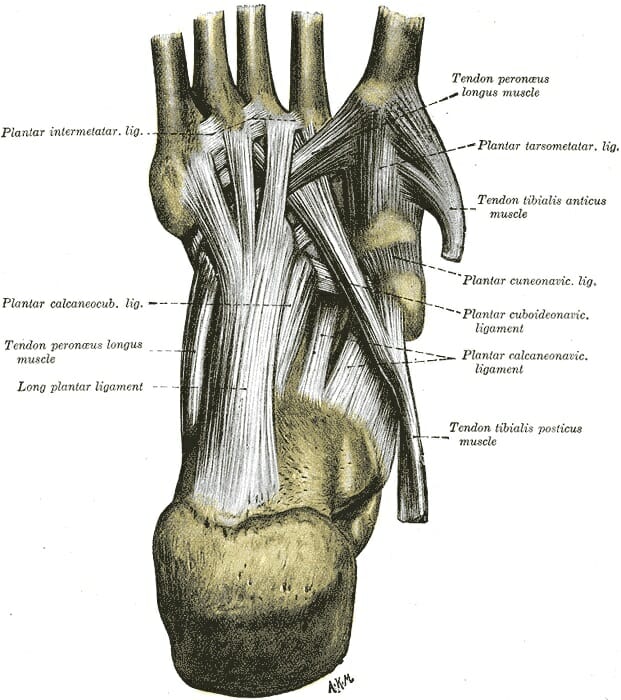
 Anatomy Of The Foot Footmaxx
Plantar Fasciitis And Bone Spurs Orthoinfo Aaos
 Feet Human Anatomy Bones Tendons Ligaments And More
 Anatomical Overlays Bottom Of The Foot These Images Will
 Notes On Anatomy And Physiology Using Imagery To Relax The
 Management Of Painful Plantar Fat Pad Atrophy Lower
 Anatomy Of Our Sandals Bay Area Custom Footwear
 Contemporary Foot Anatomy Bottom Gift Image Of Internal
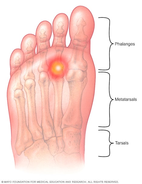 Metatarsalgia Symptoms And Causes Mayo Clinic

 Anatomy Bottom View Foot Stock Illustrations 44 Anatomy
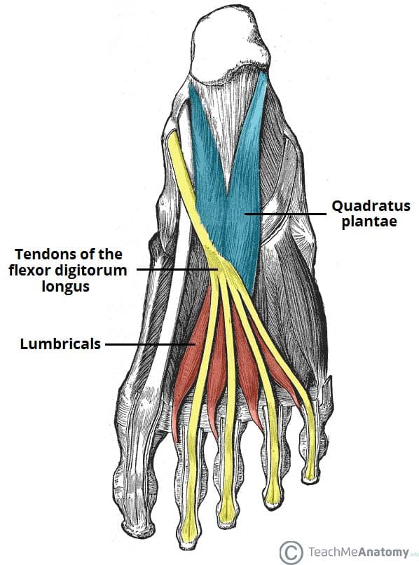
Muscles Of The Foot Dorsal Plantar Teachmeanatomy
 Foot Anatomy Eorthopod Com
 Notes On Anatomy And Physiology Using Imagery To Relax The
 Broken Ankle Types Of Fractures Diagnosis Treatments
 Plantar Fascia Wikipedia
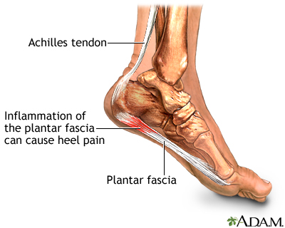
 Plantar Fasciitis Info Florida Orthopaedic Institute
 Sole Foot Wikipedia
 Common Foot And Ankle Conditions Pro Sports Orthopedics
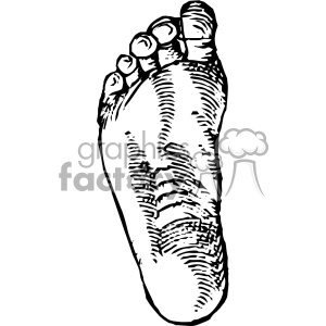 Cousin Jehan Vintage Bottom Of Foot Vector Anatomy Art Clipart Royalty Free Clipart 403137
 Plantar Fasciitis And Heel Spurs Sierra Pacific Orthopedics
 Foot Anatomy Bones Ligaments Muscles Tendons Arches
 Plantar Fasciitis Medlineplus Medical Encyclopedia
 Line Drawing Of The Left And Right Foot Soles Stock Vector
Belum ada Komentar untuk "Foot Bottom Anatomy"
Posting Komentar