Inguinal Hernia Anatomy
Direct where the peritoneal sac enters the inguinal canal though the posterior wall of the inguinal canal. These layers are a bit different between the umbilical region and the groin but overall the basic layers are the same.
This may include pain or discomfort especially with coughing exercise or bowel movements.

Inguinal hernia anatomy. It presents as a swelling above and medial to pubic tubercle above the inguinal ligament. Hernia anatomy the layers of the abdominal wall the first concept to understand is the basic layers of the abdominal wall. The 2 types of inguinal hernias are direct inguinal hernias and indirect inguinal hernias.
Often it gets worse throughout the day and improves when lying down. Clearly this definition applied to this patient. An inguinal hernia is the protrusion of intra abdominal contents through a defect in the abdominal wall.
Anatomy and management is intended for general surgeons and hernia specialists. Indirect where the peritoneal sac enters the inguinal canal through the deep inguinal ring. Giant inguinal hernia gih is defined as hernia extending below the midpoint of the inner thigh in the standing position.
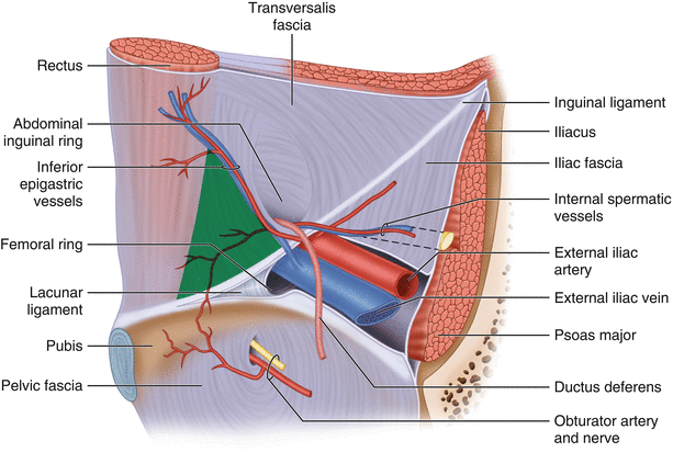
Within the boundaries of this area you can find the external iliac artery and vein. Symptoms are present in about 66 of affected people. It can be fat bowel or in some cases the genitourinary tract.
The goal of this activity is to define current treatment protocols and clinical strategies and describe state of the art materials and techniques used in the surgical management of inguinal hernias. During a laparoscopic inguinal hernia repair the dangerous triangle the triangle of doom refers to a triangular area bound by the vas deferens the testicular vessels and the peritoneal fold. Hernias involving the inguinal canal can be divided into two main categories.
The hernias were diagnosed as indirect hernias thus all of the herniated viscera passed through the inguinal canal which is unusual for his age group. An inguinal hernia is a protrusion of abdominal cavity contents through the inguinal canal. Inguinal hernia is a protrusion of loop of small intestine into the inguinal canal is termed as inguinal hernia.
Femoral branch of the genitofemoral. A 45 year old man had developed a direct inguinal hernia several months after having an emergency appendectomy. The examining doctor linked the cause of hernia to accidental nerve injury that happened during appendectomy and weakened the falx inguinalis.
Which nerve had been injured.
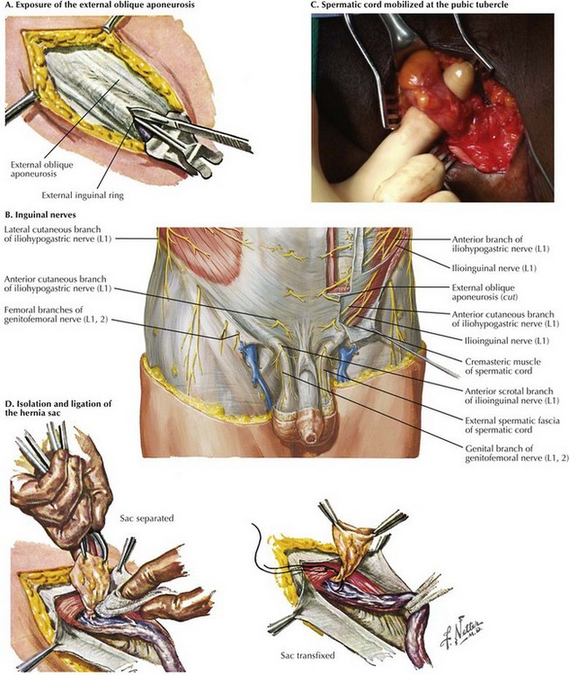 Open Inguinal Hernia Repair Basicmedical Key
 Umbilical Hernia Laparoscopic Inguinal Hernia Repair Belly
 Laparoscopic Anatomy Of Inguinal Hernia
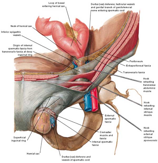 Hernia Anatomy California Hernia Specialists
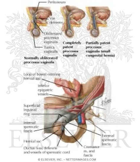 Abdominal Wall Inguinal Hernia
 Anatomy Of The Inguinal Region Simplified
 Surgical Anatomy Of Inguinal Hernia
 Indirect Inguinal Hernia Causes Symptoms Diagnosis
 Anatomy Ireland Clinical Case 030312 008 Direct Inguinal Hernia
Inguinal Hernia Cleveland Clinic
 Biodesign Inguinal Hernia Graft Cook Medical
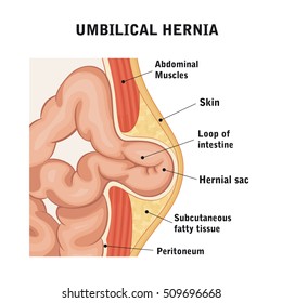 Inguinal Hernia Images Stock Photos Vectors Shutterstock
 A Triple Inguinal Hernia Right Double Indirect Hernia
 Inguinal Hernia Alvaro Garcia Md
 Inguinal Hernia In Infancy And Children Intechopen
 14 Inguinal Hernia Target Anatomy Left Titles Incision
 14 Inguinal Hernia General Anatomy Titles1 Incision Academy
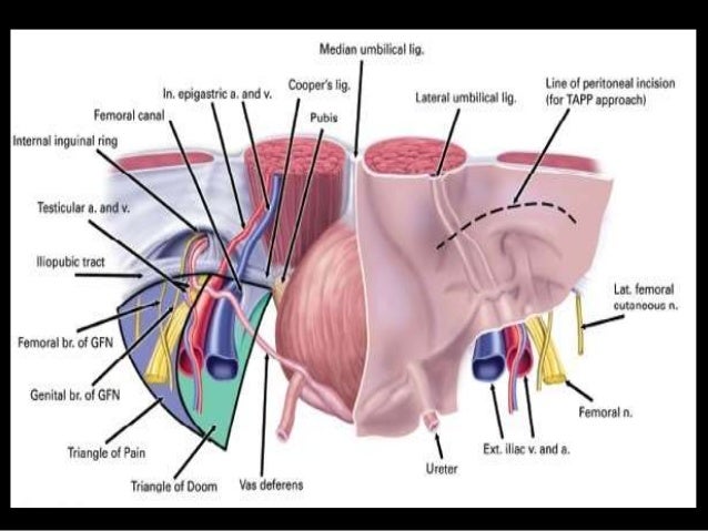 Laparoscopic Anatomy Of Inguinal Canal
 Laparoscopic Transabdominal Preperitoneal Inguinal Hernia
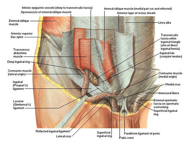 Types Of Inguinal Hernia Sports Hernia Specialist
 Inguinal Hernia Hernia Inguinal Radiology Imaging
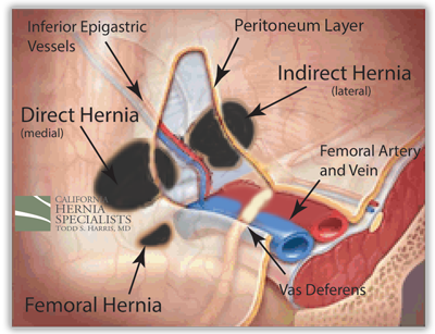 Hernia Anatomy California Hernia Specialists
 Indirect Inguinal Hernia Repair Ami 2017 Annual Conference
 Hasselbach Triangle In Relation To Inguinal Canal Medical
Anatomy Essentials For Laparoscopic Inguinal Hernia Repair


Belum ada Komentar untuk "Inguinal Hernia Anatomy"
Posting Komentar