Lung Anatomy Hilum
Hilum of the lung. This post hilum of lung belong to following categorycategories you may also find more related and detailed contents in these categories.
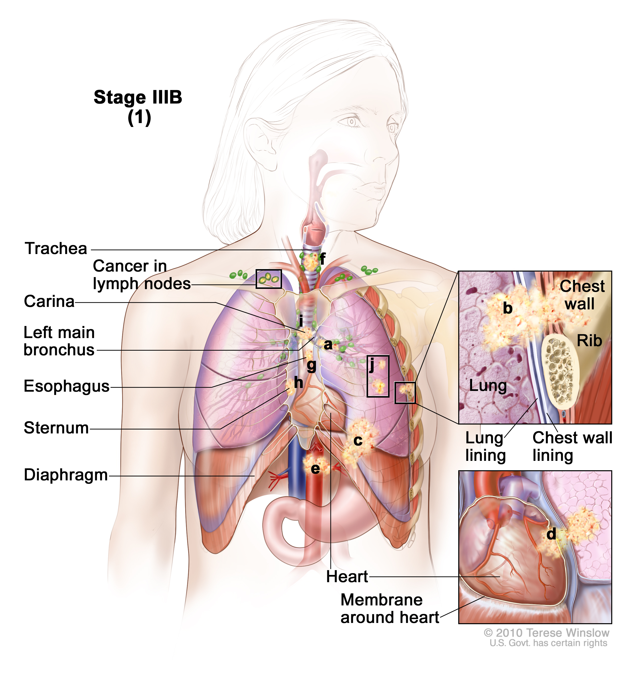 Stage Iiib Non Small Cell Lung Cancer Patient Siteman
The left and right lung roots are similar but not identical.

Lung anatomy hilum. Lymph nodes and vessels. Gross anatomy with the mediastinum at the hilum a circumscribed area where airways blood and. Both the right and the left lung have a hilum which lies roughly midway down.
The lung hilum where structures enter and leave the lung is located on this surface. The hilum of the lung is a triangular impression that allows the structures which make up the root of the lung to enter and exit. The rib cage is separated from the lung by a two layered membranous coating the pleura.
A hilum is a section of an organ where other types of structures like veins or arteries can enter. Gross anatomy left hilum. The hilum of the lung is a wedge shaped section in the central area.
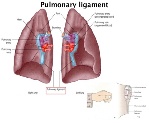
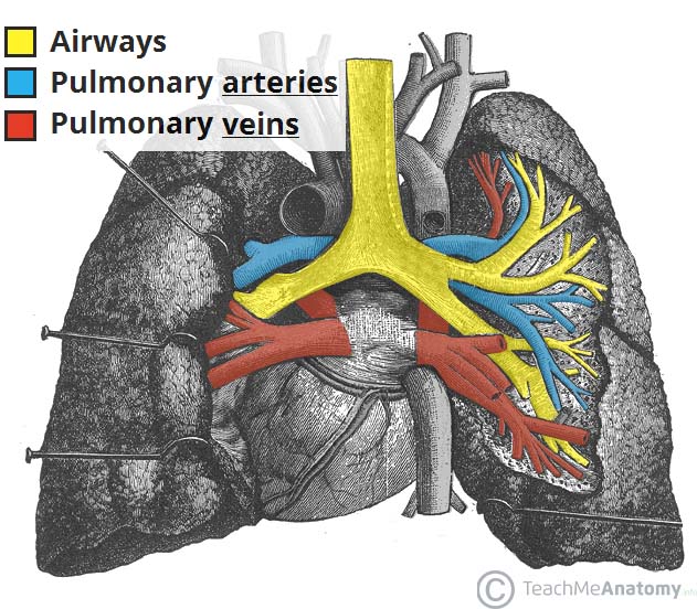
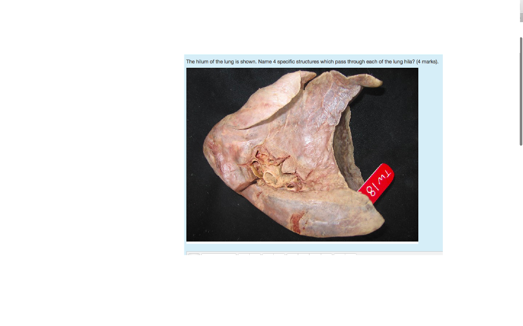
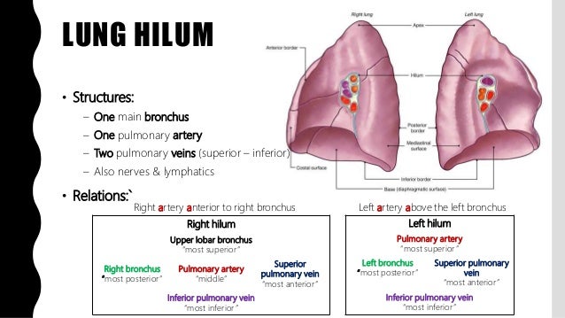
The following structures are found at each hilum. Lung root consists of the structures passing to and from the hilum of the lung to the mediastinum. The hilar region of.
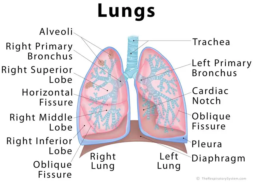
It is nearer to the back than the front. Respiratory system anatomy hyoid bone larynx and thyroid gland location in real human. The base of the lung is formed by the diaphragmatic surface.
Structure of lung in lung to its apex is the hilum the point at which the bronchi pulmonary arteries and veins lymphatic vessels and nerves enter the lung. Tests to evaluate the hilum. Lung roots lie opposite to t5 t7 vertebrae.
In human respiratory system. The root of the lung is connected by the structures that form it to the heart and the trachea. This concavity is deeper in the right lung due to the higher position of the right dome overlying the liver.
The root of the lung is located at the hilum of each lung just above the middle of the mediastinal surface and behind the cardiac impression of the lung. Abnormalities in the hilum are usually noted on imaging. Anatomy and abnormalities anatomy of the hilum.
Below this is the left main bronchus. There are two pulmonary veins one lies in front and the other lies below the left main bronchus. The structures of the lung root are embedded in the connective tissue and surrounded by extension of mediastinal pleura.
In the left hilum the left pulmonary artery occupies the upper part. It rests on the dome of the diaphragm and has a concave shape. The hilum is the large triangular depression where the connection between the parietal pleura and the visceral pleura is.
 Pulmonary Cavities Anatomy An Essential Textbook 1st Ed
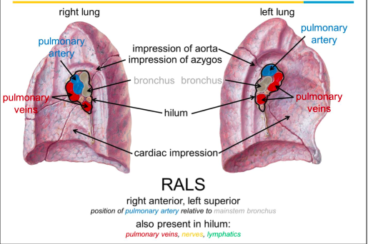
 Anatomy Of The Lung Flashcards Quizlet
 Anatomy Of Lung Hilum Lung Anatomy Heart Anatomy

/iStock_000006469946_Large-56a5c5575f9b58b7d0de6a59.jpg) Hilum Of The Lung Definition Anatomy And Masses
Hilum Of The Lung Definition Anatomy And Masses
 Difference Between Hilum And Root Of Lung Compare The
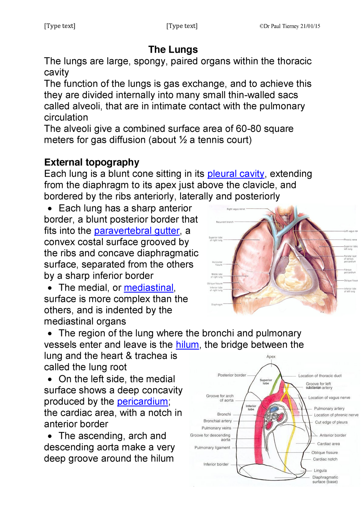 Anatomy Of The Lungs Pleura Anat10110 Ucd Studocu
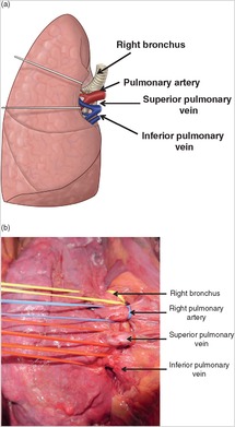 Lung Injuries Chapter 17 Atlas Of Surgical Techniques In
 The Lungs Position Structure Teachmeanatomy
Summary Netter S Anatomy Lecture Lungs Dbs 8110 Studocu
 What Is The Hilum Of The Lung Youtube
 Lungs The Big Picture Gross Anatomy 2e Accessmedicine
 Solved The Hilum Of The Lung Is Shown Name 4 Specific St
 Lung Cancer The Patient Guide To Heart Lung And
 Lung Injuries Chapter 17 Atlas Of Surgical Techniques In
 Lungs Definition Location Anatomy Function Diagram




Belum ada Komentar untuk "Lung Anatomy Hilum"
Posting Komentar