Sacroiliac Joint Anatomy
They also form a strong base for the spine. Movement at the sacroiliac joint is minimal and is limited to gliding and rotation.

The si joint is stabilized by a network of ligaments and muscles which also limit motion.

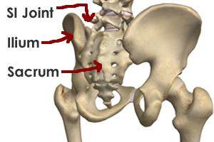
Sacroiliac joint anatomy. Commonly called the si joint the sacroiliac joint is really a pair of joints that function in concert with one another. Amongst other reasons because the sij surfaces are covered by two different kinds of cartilage. The sacroiliac joint allows for the slight shifting of these bones relative to each other to increase the flexibility of the pelvis especially during childbirth.
Sacrotuberous ligament this is a triangular shaped flat ligament that runs from. Si joint anatomy and function. Sacrospinous ligament this is another thin triangular shaped ligament of.
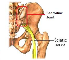
The normal sacroiliac joint has a small amount of normal motion of approximately 2 4 mm of movement in any direction. The sacroiliac joint lies between the sacrum and the ilium. Sacroiliac joint si joint pain typically results in pain on one side very low in the back or in the buttocks.
The bodys shock absorber. The si joint may be a source of low back pain however any structure in the lower back. Sacroiliac joint anatomy sacroiliac joint anatomy takes you on a little tour of the pelvis.
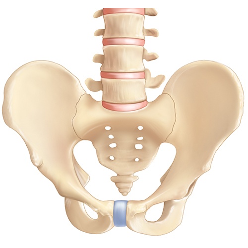
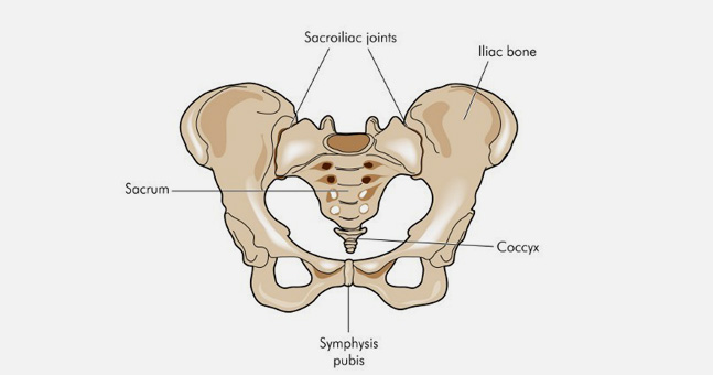
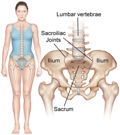
The sacrum lies at the lower part of the spine. The sacroiliac joint is a synovial planar joint formed in the pelvis between the ilium and the sacrum. Other ligaments of the sacroiliac joint iliolumbar ligament this ligament extends from the lateral surface of the transverse process.
Being a synovial joint it is surrounded by a capsule. Another term for sacroiliac joint pain is sacroiliitis a term that describes inflammation in the joint. An overview of its anatomy function and potential clinical implications a vleeming 1 2 m d schuenke 1 a t masi 3 j e carreiro 1 l danneels 2 and f h willard 1 1 department of anatomy university of new england college of osteopathic medicine biddeford me usa.
The sacroiliac joint is formed by the irregularly shaped auricular surfaces. The ligaments of the sacroiliac joint include the following. The left and right sacroiliac joints support and transmit the weight of the body to the legs through the pelvis.
This is quite unlike any other part in the body.
 Minimally Invasive Sacroiliac Joint Fusion Globus Medical
 Sacroiliac Joint Dysfunction A Crucial Element Of
 Sacroiliac Joint Pain Syndrome Si Joint Pain Vail Aspen
 Anatomy Of The Sacroiliac Joint Human Anatomy
 Sacroiliac Joint Dysfunction And Piriformis Syndrome The
 Sacroiliac Joint Is Different From Other Joints
:max_bytes(150000):strip_icc()/GettyImages-87394799-56a05fac5f9b58eba4b0275a.jpg) Sacroiliac Joint Anatomy And Characteristics
Sacroiliac Joint Anatomy And Characteristics

 The Anatomy Of The Si Joint And Its Relationship To Yoga
 Posterior Pelvic Ring Fractures Of The Si Joint And The
 Nutation Counternutation Yoga Anatomy
 What Are The Causes Of Sacroiliac Joint Dysfunction
Sacroiliac Joint Pain Western New York Urology Associates Llc

 Sacroiliac Joint Dysfunction Treatment Symptoms Pain Relief
 The Anatomy Of The Si Joint And Its Relationship To Yoga
 Orthoneurospine And Pain Institute Sacroiliac Joint
 Where Is The Sacroiliac Joint Anatomy Of The Sacroiliac Joint
 Sacroiliac Ligaments Si Joint Pain Serola Biomechanics Inc
 Sacroiliac Joint Injections Dr Edward Magaziner Nj
 Standing Flexion Test Physiopedia
Anatomy Hip And Sacroiliac Joint Anat10110 Ucd Studocu
 The Sacroiliac Joint 5 Things We Didn T Learn In Yoga
 The Importance Of Si Joint Function Phila Massages
 What Is The Si Joint Si Joint Anatomy Si Bone
 Sacroiliac Joint Montgomery Spine Center
 Protect Your Yoga Students Sacroiliac Joints Yoga
 Sacroiliac Joint Syndrome Redlands Loma Linda Highland
 Anatomy Of The Sacroiliac Joint Active Family Chiropractic
The Daily Bandha The Sacroiliac Joint

Belum ada Komentar untuk "Sacroiliac Joint Anatomy"
Posting Komentar