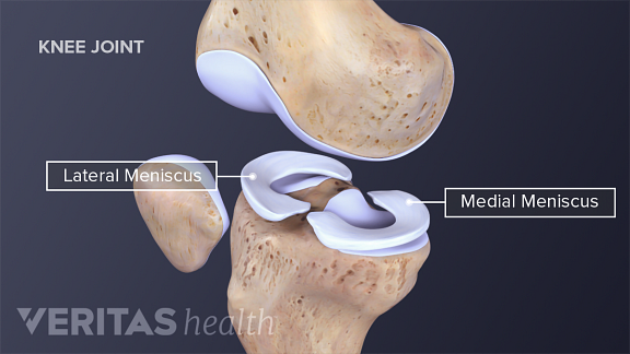
Meniscus Knee Anatomy
Any activity that causes you to forcefully twist or rotate your knee especially when putting your full weight on it can lead to a torn meniscus. However anyone at any age can tear a meniscus.
 Meniscal Anatomy In Radiology
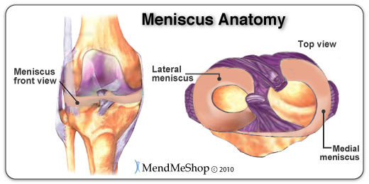
They are concave on the top and flat on the bottom articulating with the tibia.

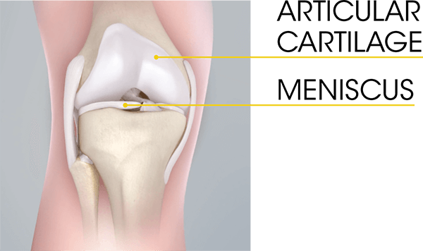
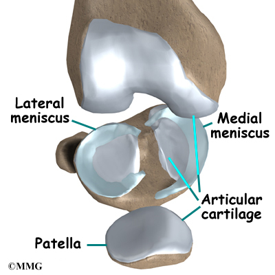
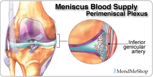
Meniscus knee anatomy. It acts like a hinge allowing the knee to flex bend and extend straighten. Collection of fluid in the back of the knee. In most of our joints including the knee there is a layer of articular cartilage which is made of collagen and chondroitin.
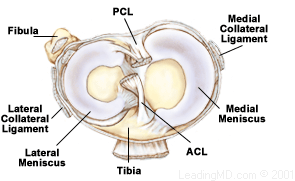
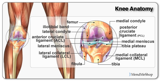
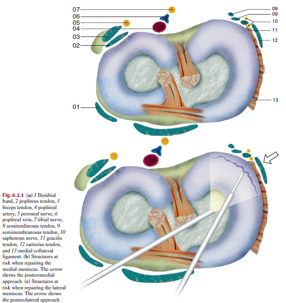
Their incidence like many orthopaedic ailments increases with age. The knee joint contains the meniscus structure comprised of both a medial and a lateral component situated between the corresponding femoral condyle and tibial plateau figure 1. The knee is the largest joint in the body.
Meniscus tears are among the most common knee injuries. The knee is a joint where the bone of the thigh femur meets the shinbone of the leg tibia. Each is a glossy white complex tissue comprised of cells specialized extracellular matrix ecm molecules and region specific innervation and vascularization.
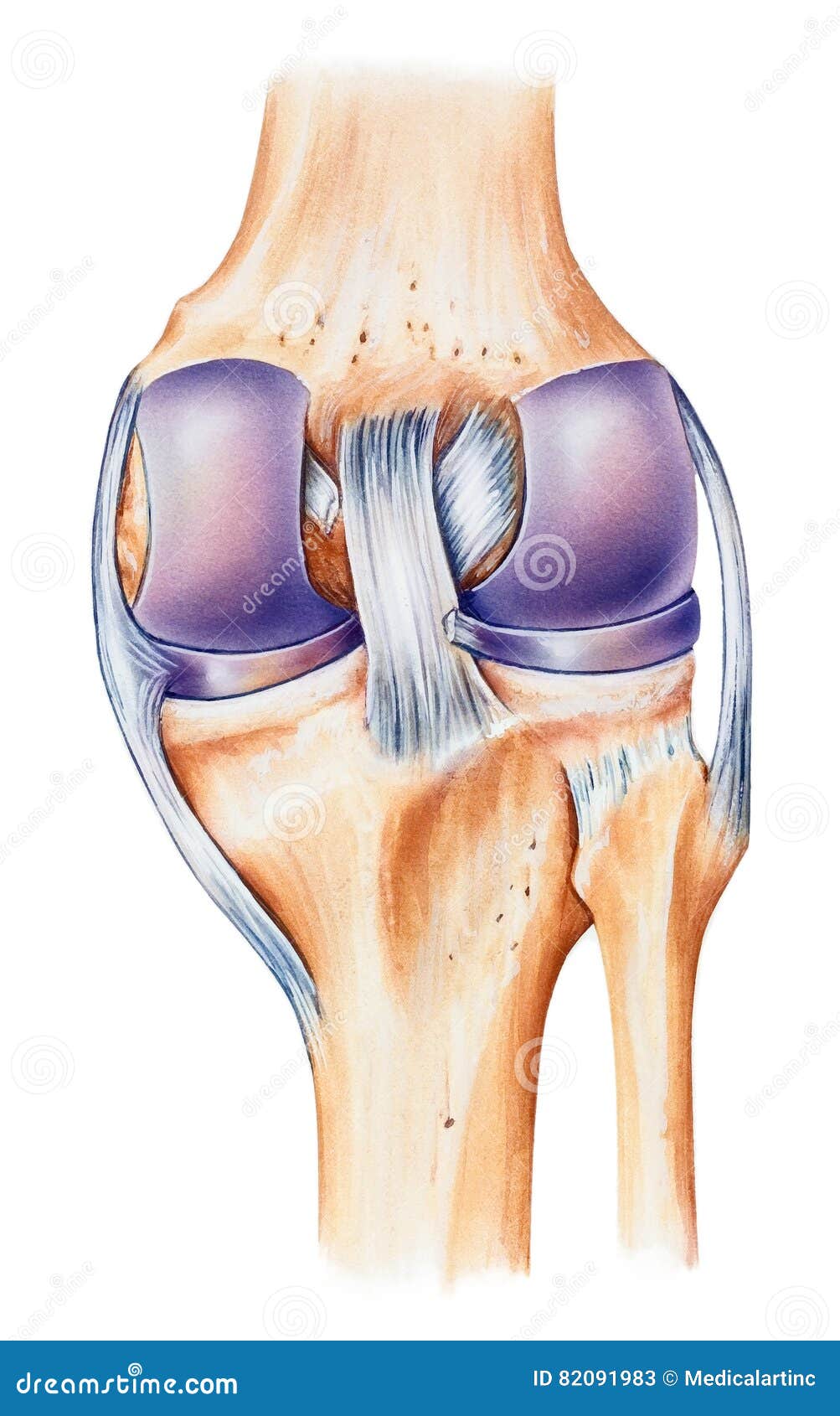
The band goes around the knee joint in a crescent shaped path and is located between the medial condyles of the shin and the femur or thighbone. The medial meniscus is the central band of cartilage attached to the tibia or shinbone. There are four ligaments of the joint the medial and lateral collateral ligaments and.
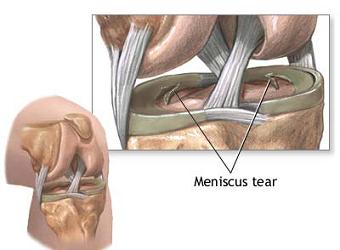
It provides a smooth surface over the bones. Meniscal tears can result from nearly any activity involving bending or twisting of the knee. Their main role is shock absorption improve stability of the knee joint and load transmission.
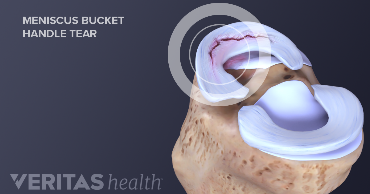
Picture of torn meniscus. Its job is to cushion the joint and transfer forces between the tibia and femur bones. The knee menisci are fibrocartilaginous structures that sit within the knee joint deepening the tibiofemoral articulation.
Meniscus anatomy the menisci of the knee are two pads of fibrocartilaginous tissue which serve to disperse friction in the knee joint between the lower leg tibia and the thigh femur. When people talk about torn cartilage in the knee they are usually referring to a torn meniscus. They are attached to the small depressions fossae.
Athletes particularly those who play contact sports are at risk for meniscus tears. Pain swelling and warmth in any of the bursae of the knee. The medial condyles are areas of these bones located on the inner sides of the knees.
They often occur while performing athletics but can also frequently occur during nonathletic activities. Bursitis often occurs from overuse or injury. Each of your knees has two c shaped pieces of cartilage that act like a cushion between your shinbone and your thighbone menisci.
Torn meniscus anatomy and causes video a torn meniscus is one of the most common causes of knee pain. The knee meniscus is a special layer of extra cartilage that lines the knee joint. A torn meniscus is one of the most common knee injuries.
 Lateral Meniscus Wikipedia
 Physical Therapy To Treat Torn Meniscus Comparable To
 Understanding Meniscus Tears
 Figure Anatomy Of The Right Knee Download Scientific Diagram
 Anatomy Of The Knee Central Coast Orthopedic Medical Group
 Clinical Anatomy Knee Mensicus And Knee Joint
The Knee Meniscus Structure Function Pathophysiology
 Meniscus Injuries
Common Knee Injuries Orthoinfo Aaos
 Pin On Yoga
Knee Anatomy
 Understanding The Role Of Cartilage In The Knee
 Atro Medical Meniscus Vervanging Replacement Atro Medical
 Torn Meniscus Symptoms Treatment Mri Test Recovery Time
 Anomy Of Knee Iron Thumb
 Acl Solutions Acl Knee Anatomy And Diagram Images
Meniscus Tears Orthoinfo Aaos
 Understanding Meniscus Tears
 The Knee Bellin Orthopedic Surgery Center

 Meniscal Repair Physiopedia
 Meniscus Anatomy Wikipedia
 Knee Anatomy Dorsal View Stock Illustration
Belum ada Komentar untuk "Meniscus Knee Anatomy"
Posting Komentar