Microscopic Anatomy Of Liver
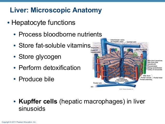
It functions as a phagocyte a cell that engulfs and destroys foreign material or other cells. At the porta hepatis the glisson capsule travels along the portal tracts.
 Liver Anatomy Overview Gross Anatomy Microscopic Anatomy
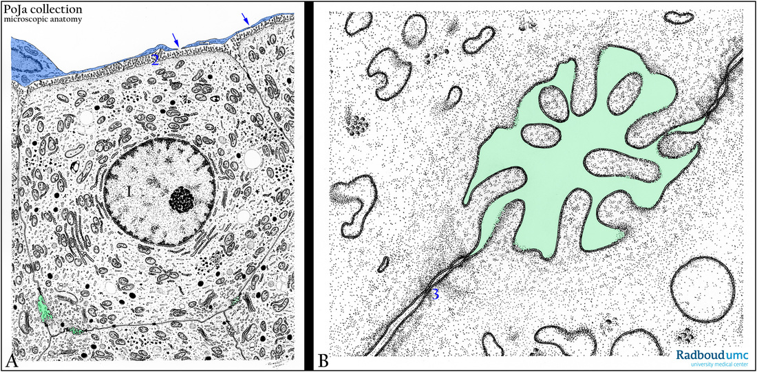
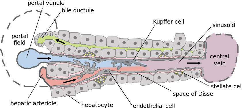
Hepatocytes have microvilli that extend into space.

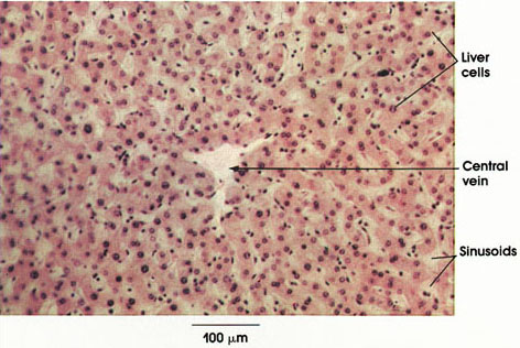
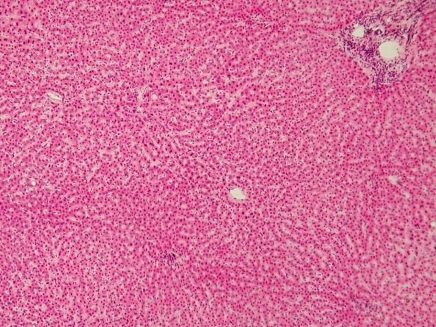
Microscopic anatomy of liver. Such structural and functional organization allows assessment of diffuse disease processes in small representative biopsy specimens. Microscopic anatomy the study of microscopic anatomy shows two major types of liver cell. Anatomically the liver consists of four lobes.
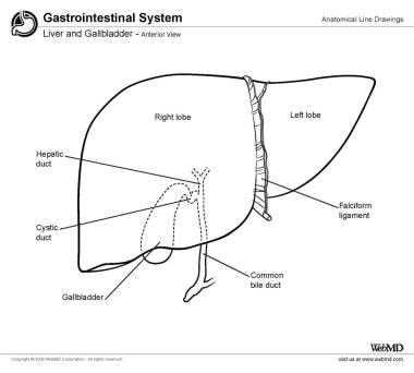
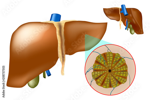
The liver is a complex three dimensional structure that consists of epithelial and mesenchymal elements arranged in repetitive microscopic units. Mononuclear phagocytic system destroy erythrocytes digest hemoglobin and destroy bacteria. Two larger ones right and left and two smaller ones quadrate and caudate.
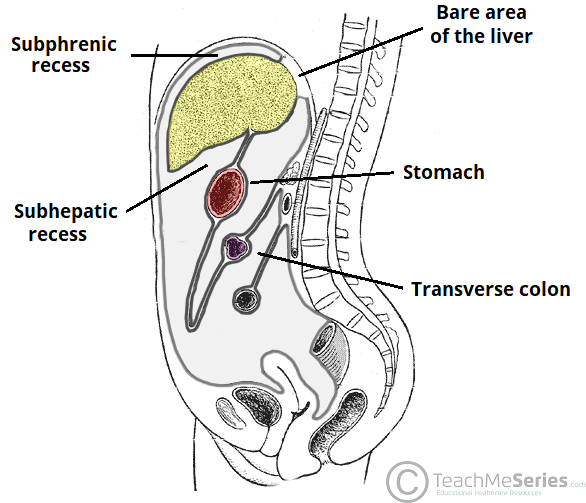
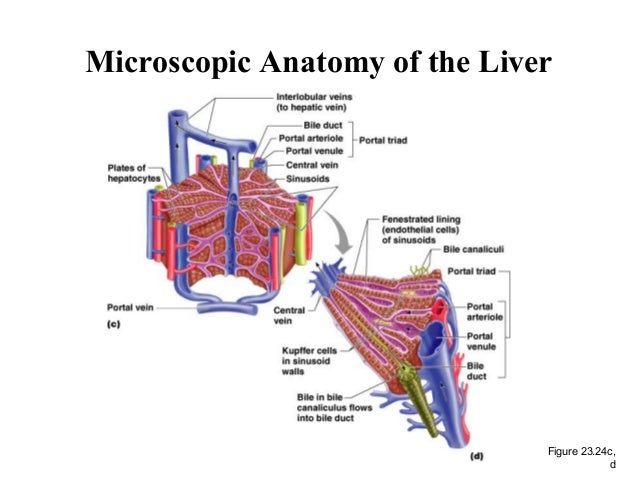
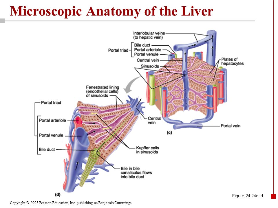
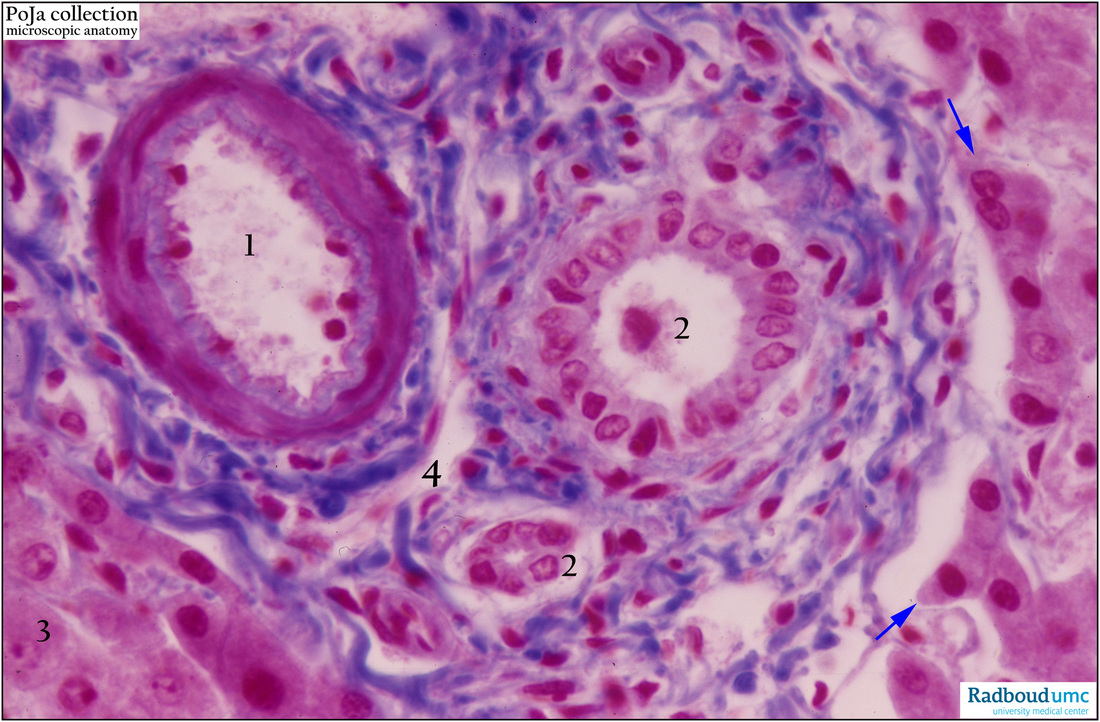
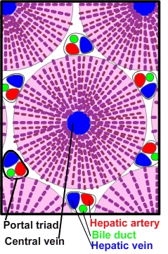
The macroscopic and microscopic anatomy of the liver is difficult to understand partly because of its inherently complicated three dimensional structure and partly because of the recent trend to replace simple but misleading morphological descriptions with more accurate but less obvious functional descriptions. Microscopic anatomy the surface of the liver is covered by visceral peritoneum serosa with a glisson capsule underneath. The other major cell of the liver the kupffer cell adheres to the wall of the sinusoid and projects into its lumen.
Nonparenchymal cells constitute 40 of the total number of liver cells but only 65 of its volume. Histology the study of microscopic anatomy shows two major types of liver cell. Small spaces disse spaces are present in places between the hepatocyte and the sinusoidal endothelium.
Anatomy of the liver. 7085 of the liver volume is occupied by parenchymal hepatocytes. Such structural and functional organization allows assessment of diffuse disease processes in small representative biopsy specimens.
Non parenchymal cells constitute 40 of the total number of liver cells but only 65 of its volume. Histologically speaking it has a complex microscopic structure that can be viewed from several different angles. Make up 15 of liver population perisinusoidal space site of exchange of material between blood and liver cells.
Part of wall of sinusoids. Parenchymal cells and non parenchymal cells. Microscopic anatomy of the liver murli krishna md.
About 7085 of the liver volume is occupied by parenchymal hepatocytes. Parenchymal cells and nonparenchymal cells. The liver is a complex threedimensional structure that consists of epithelial and mesenchymal elements arranged in repetitive microscopic units.
Organs And Structures Of The Respiratory System Anatomy
 Anp1107 Notes Midterm 2 Pdf Anp1107 Notes The Digestive
 Medical Health Information Liver Microscopic Anatomy

 The Liver Lobes Ligaments Vasculature Teachmeanatomy
 Digestive System Full
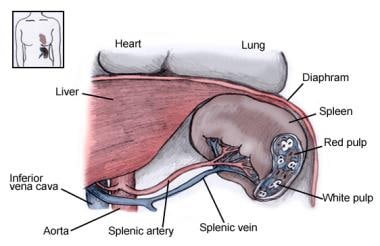 Spleen Anatomy Overview Gross Anatomy Microscopic Anatomy
 Chapter 24 The Digestive System Part E Ppt Video Online
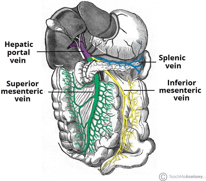 The Liver Lobes Ligaments Vasculature Teachmeanatomy
 Liver Wikipedia
Microscopic Anatomy Of Liver Youtube


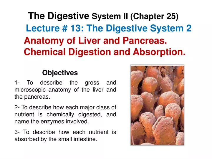 Ppt Lecture 13 The Digestive System 2 Powerpoint
 Histology The Study Of The Microscopic Anatomy Of Cells And
 Anatomy Atlases Atlas Of Microscopic Anatomy Section 1 Cells
 Microscopic Anatomy Of The Liver Hepatic Lobules The
 Microscopic Anatomy Of Liver Diagram Quizlet
 The Liver Anatomy Functions And Diseases Medical Library
 File 2423 Microscopic Anatomy Of Liver Jpg Wikipedia
 Liver Wikiwand
 Digest Ii Online
 Anatomy And Physiology Hepatitisliver
 Microscopic Anatomy Of The Liver Diagram Quizlet
 Special Report On Diseases Of Cattle Cattle Diseases Of
Belum ada Komentar untuk "Microscopic Anatomy Of Liver"
Posting Komentar