Plexus In Anatomy
It also receives contributions from thoracic spinal nerve 12. A combination of interlaced parts.
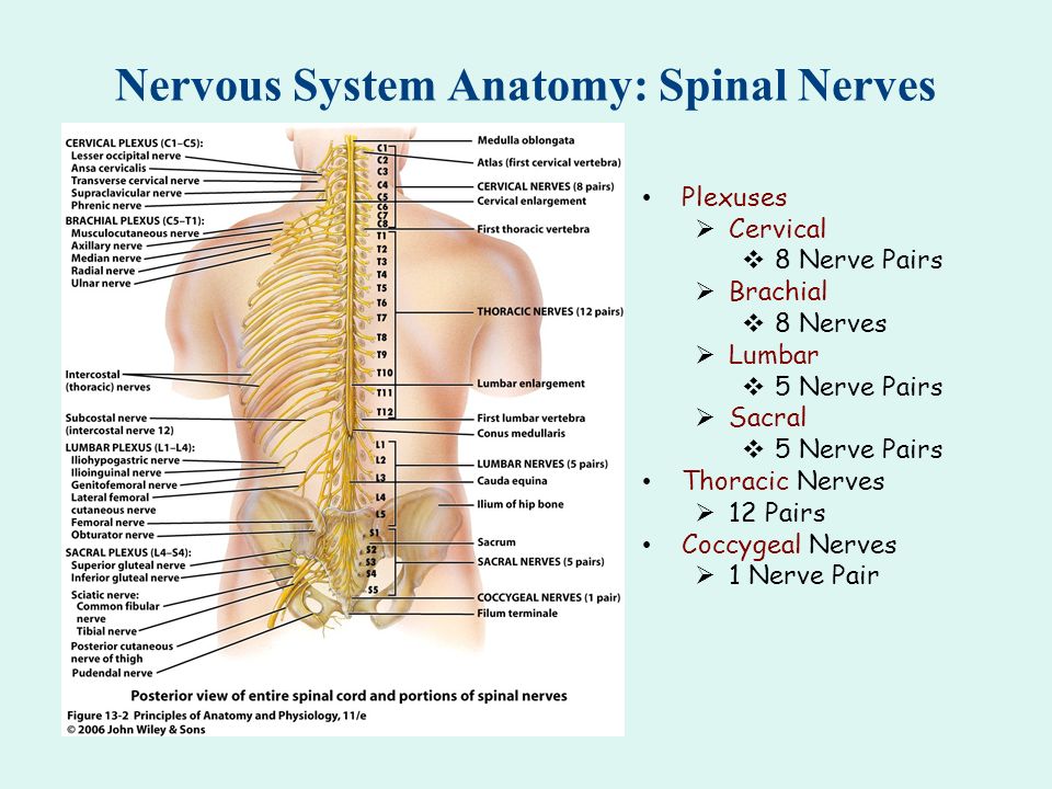 Nervous System Anatomy Neuron Ppt Download
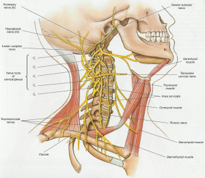
You have two cervical plexione on the left and one on the right.

Plexus in anatomy. This plexus lies within the psoas major muscle. The celiac plexus consists of celiac superior mesenteric and renal ganglia found surrounding the roots of the celiac trunk superior mesenteric and renal arteries. A nerve plexus is a plexus branching network of intersecting nerves.
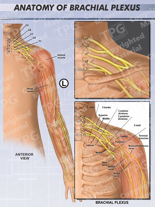
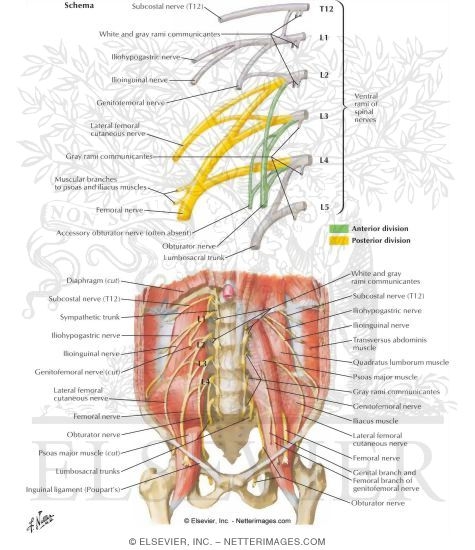
The plexus is formed by the anterior rami divisions of cervical spinal nerves c5 c6 c7 and c8 and the first thoracic spinal nerve t1. There is 1 small error the lesser occipital nerve comes from c2 only not c2 and c3. The lumbar plexus is formed by the ventral rami of l1l5 spinal nerves with a contribution of t12 form the lumbar plexus.
The cervical plexus includes six large nerves that divide into smaller branches. Thus the solar plexus is a location where a number of nerve endings meet which increases the sensitivity and functionality of this specific region. In this article we shall look at the anatomy of the lumbar plexus its formation and major branches.
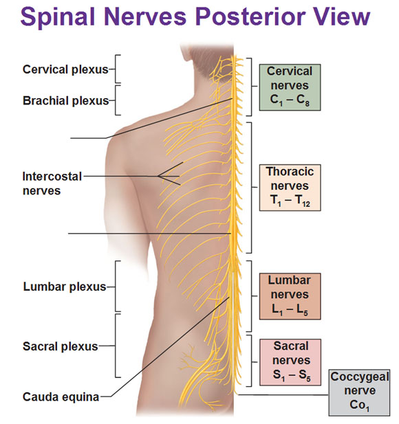
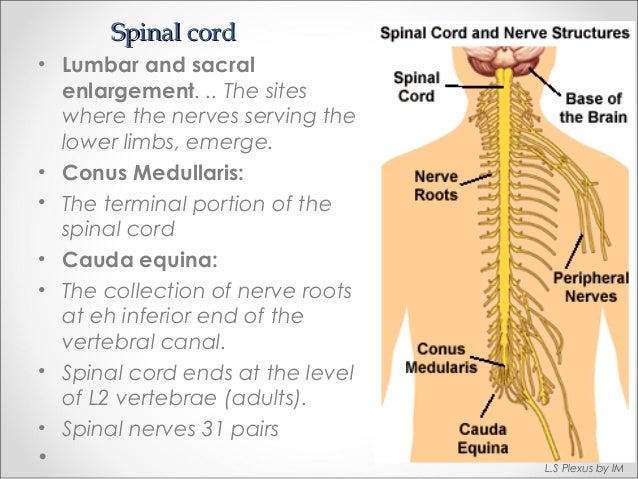
In the human body a complex collection of nerves of the nervous system in one specific location is known as a plexus. The sacral plexus or parts of the sacral plexus can be affected by. A structure in the form of a network especially of nerves blood vessels or lymphatics.
Sacral plexusserves the pelvis buttocks genitals thighs calves and feet. A nerve plexus is composed of afferent and efferent fibers that arise from the merging of the anterior rami of spinal nerves and blood vessels. The sacral plexus is formed by the lowest lumbar spinal nerves l4 and l5.
An overview of the sacral plexus anatomy. For their paraaortic location these ganglia are also called prevertebral paraaortic ganglia. The sacral plexus has extensive functions throughout the pelvis and legs.
The sacral plexus is formed by the ventral rami of l4 s3 with parts of the l4 and s4 spinal nerves. The plexus is formed by the anterior rami divisions of the lumbar spinal nerves l1 l2 l3 and l4. The left and right cervical plexi are symmetrical and should be exactly the same.
In this article we shall look at the anatomy of the brachial plexus its formation and anatomical course through the body. This is a quick way to draw out the cervical plexus.
 Sacral Nerve Plexus Human Anatomy Organs
 The Lumbosacral Plexus Human Anatomy
 Peripheral Nervous System Spinal Nerves And Plexuses
 Brachial Plexus Anatomy Order
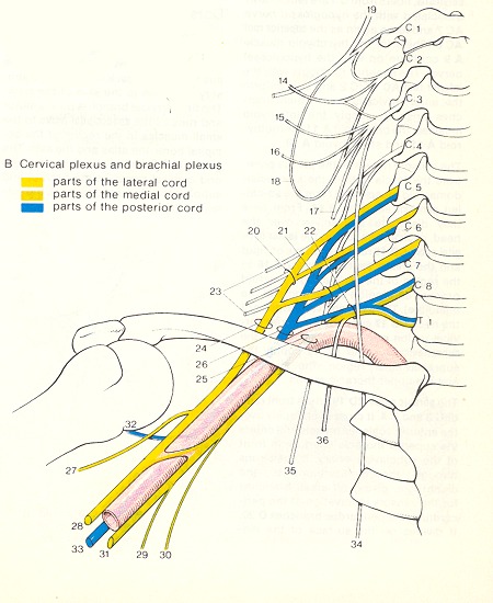
 Cervical Plexus Physiopedia
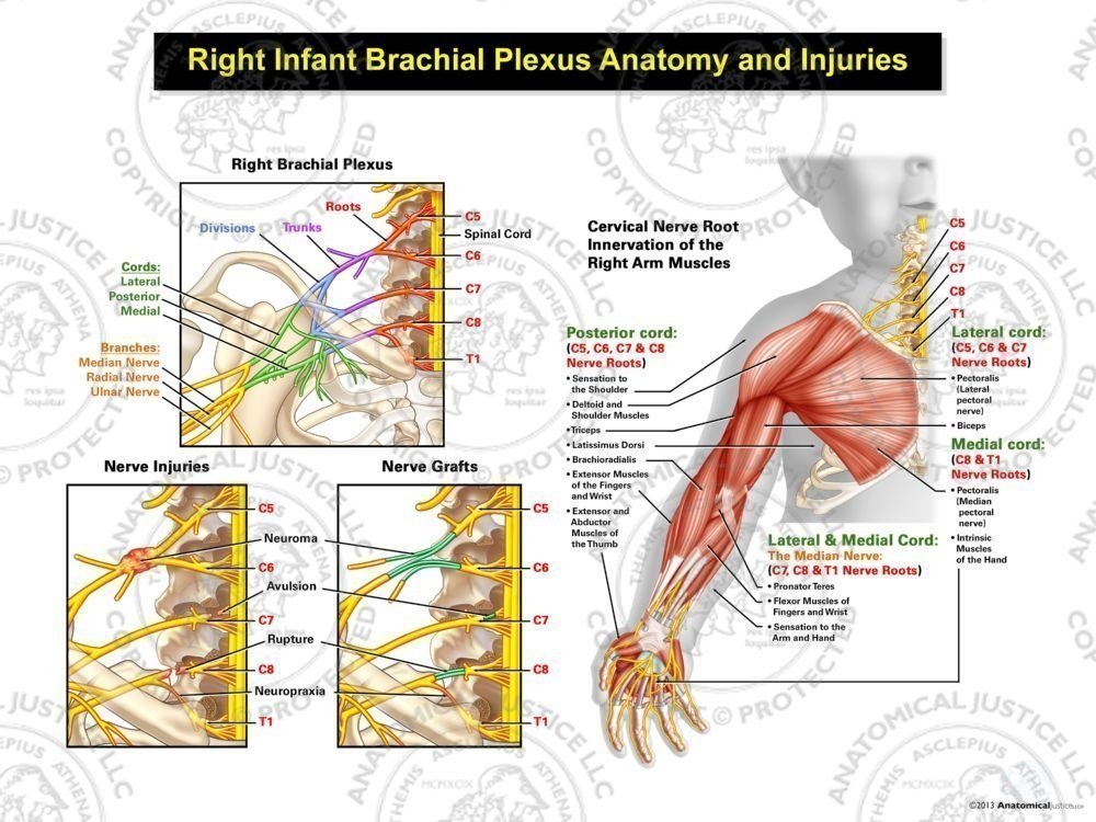 Right Infant Brachial Plexus Anatomy And Injuries
 Lumbar Plexus Block Landmarks And Nerve Stimulator
 Lumbar Plexus Nerves Lumbar Plexus
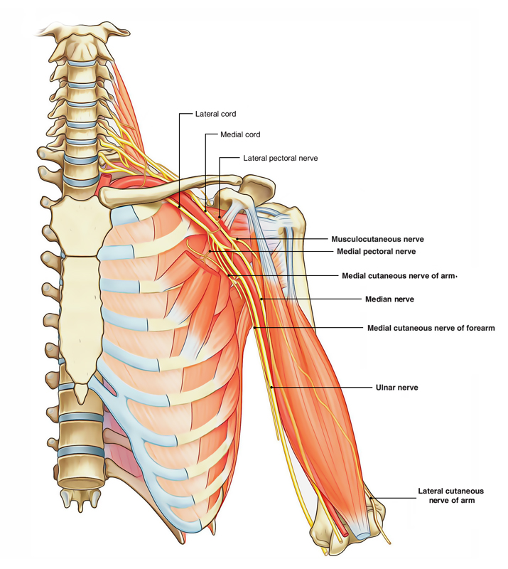
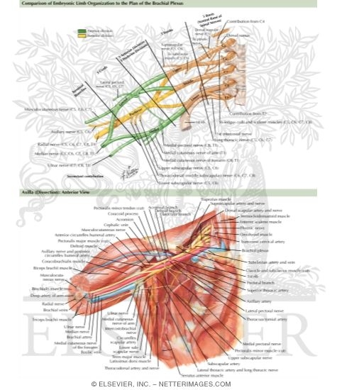 Anatomy Of Brachial Plexus
 Anatomy Of Lumbosacral Plexus By Murtaza Syed
 Lower Extremity Peripheral Nerve Blocks Lumbar Sacral
 Neonatal Pediatric Brachial Plexus Brachial Plexus
 Brachial Plexus Part 1 Anatomical Relations Sketchy Medicine
 Lumbar And Sacral Plexus
 An Overview Of The Lumbar Plexus Medone Thieme
 Anatomy And Innervation Of The Lumbar Plexus
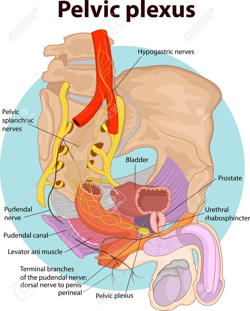 Vector Illustration Of Pelvic Plexus Anatomy
![]() Celiac Solar Plexus Definition Anatomy And Function
Celiac Solar Plexus Definition Anatomy And Function
 Untitled
 Accessphysiotherapy Lumbar And Sacral Plexus With Clinical
 Sacral Plexus Made Easy Preview Human Anatomy Kenhub
 Easy Notes On Brachial Plexus Learn In Just 4 Minutes
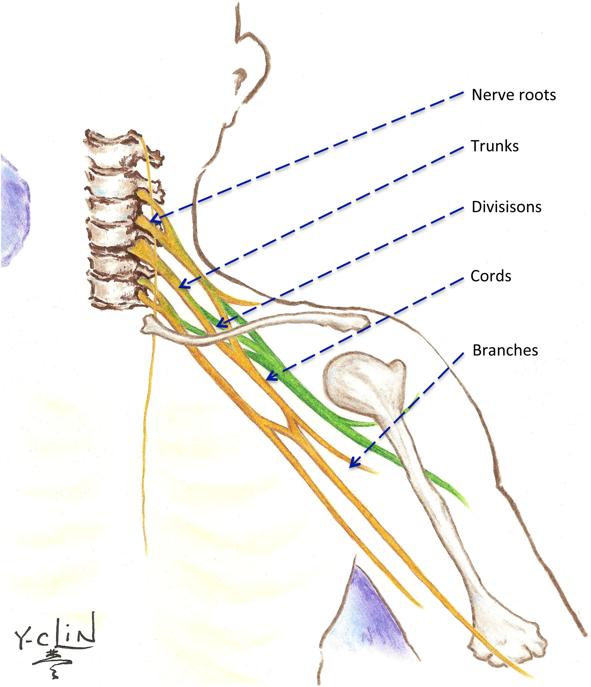
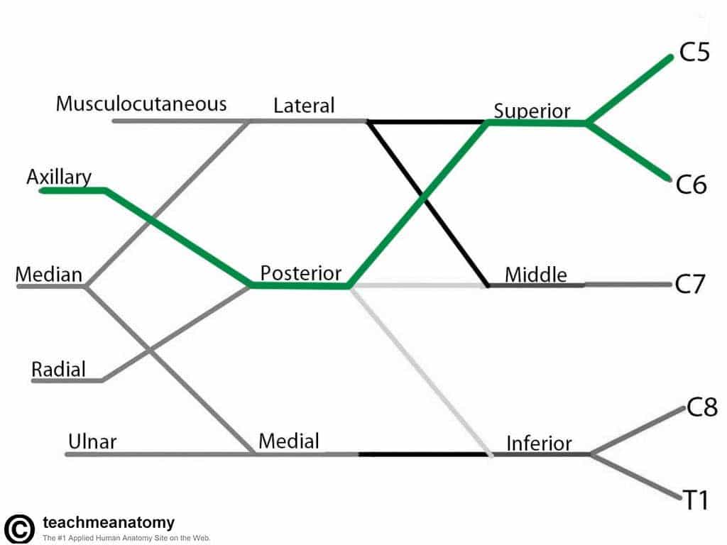 The Brachial Plexus Sections Branches Teachmeanatomy
 Anatomy And Landmarks For Branches Of The Brachial Plexus A
 1 Brachial Plexus Anatomy Image Published Under License
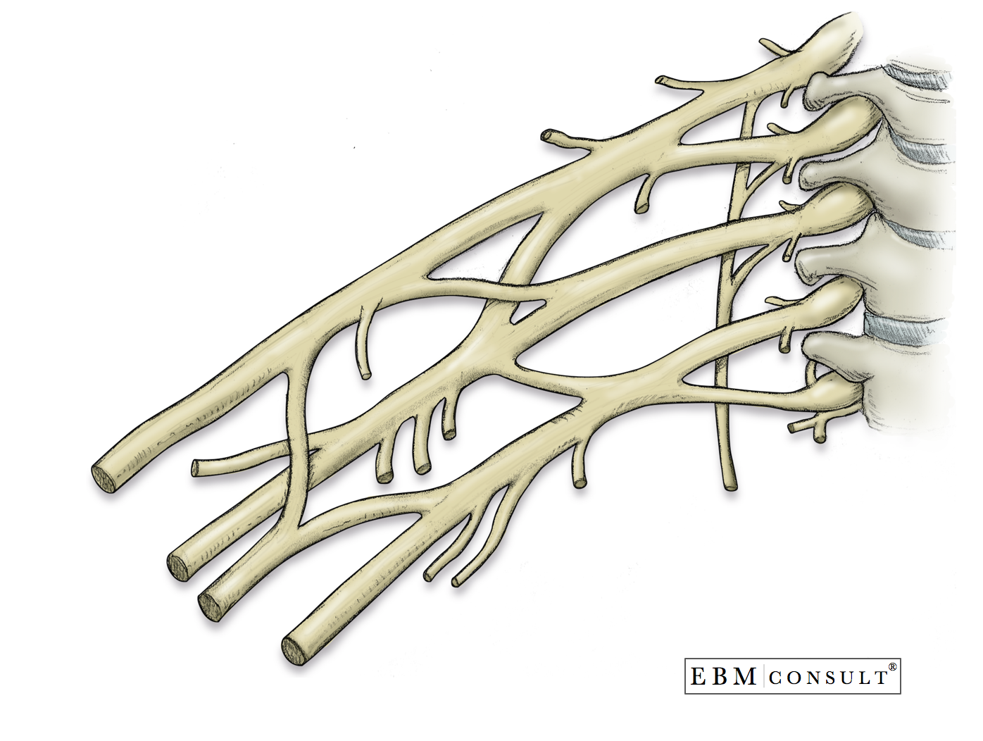
 4 Anatomy Of The Lumbosacral Plexus Image By Springer
 Anatomy Brachial Plexus
 3 Anatomy Of The Lumbar Plexus Nerves Image By Springer


Belum ada Komentar untuk "Plexus In Anatomy"
Posting Komentar