Venous Anatomy Arm
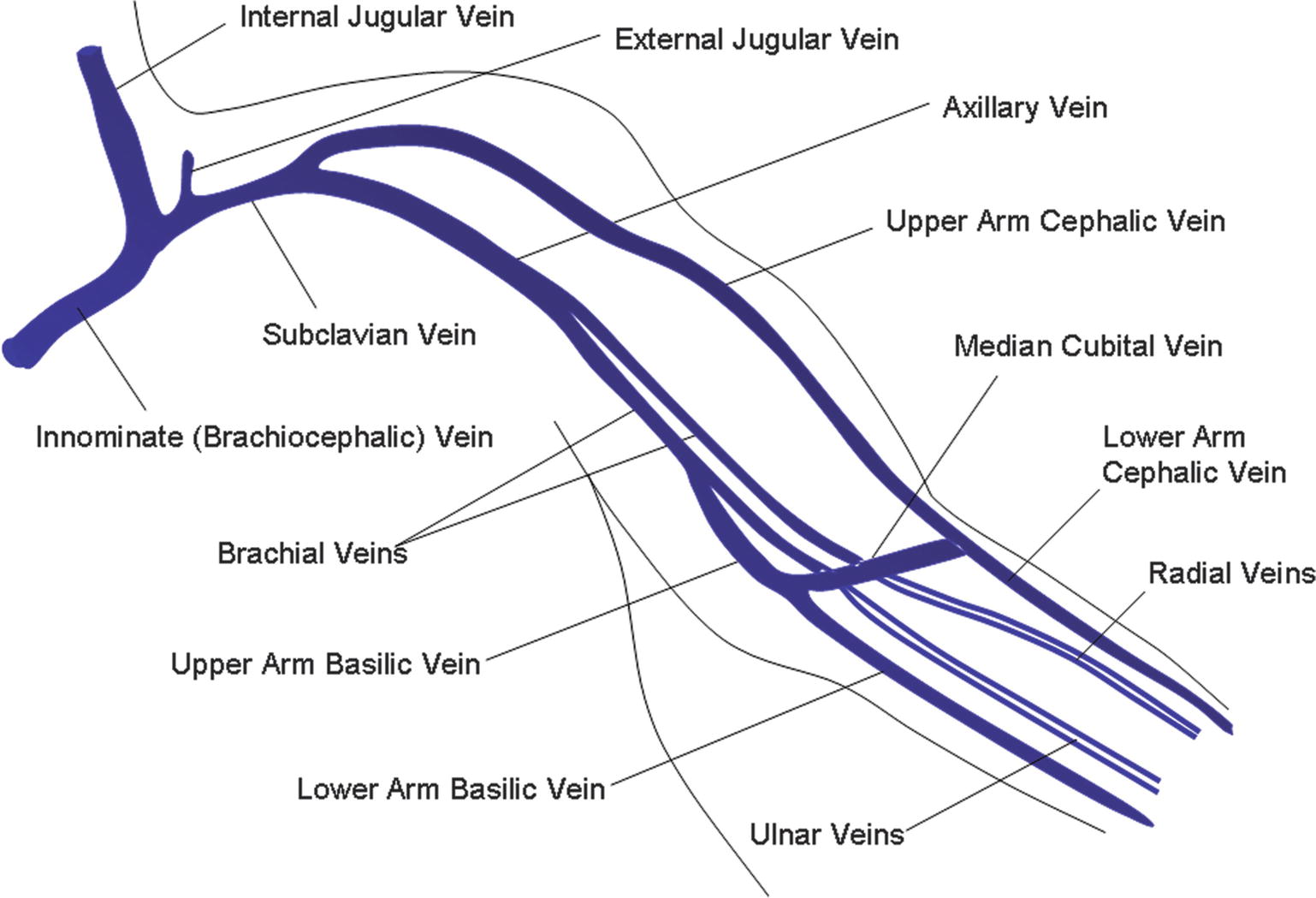
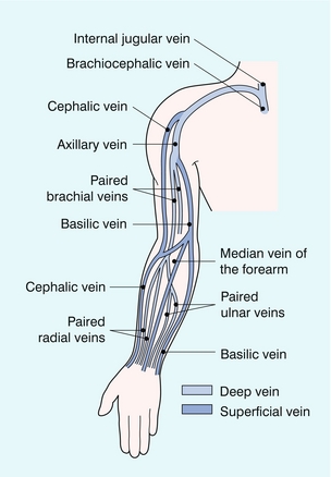
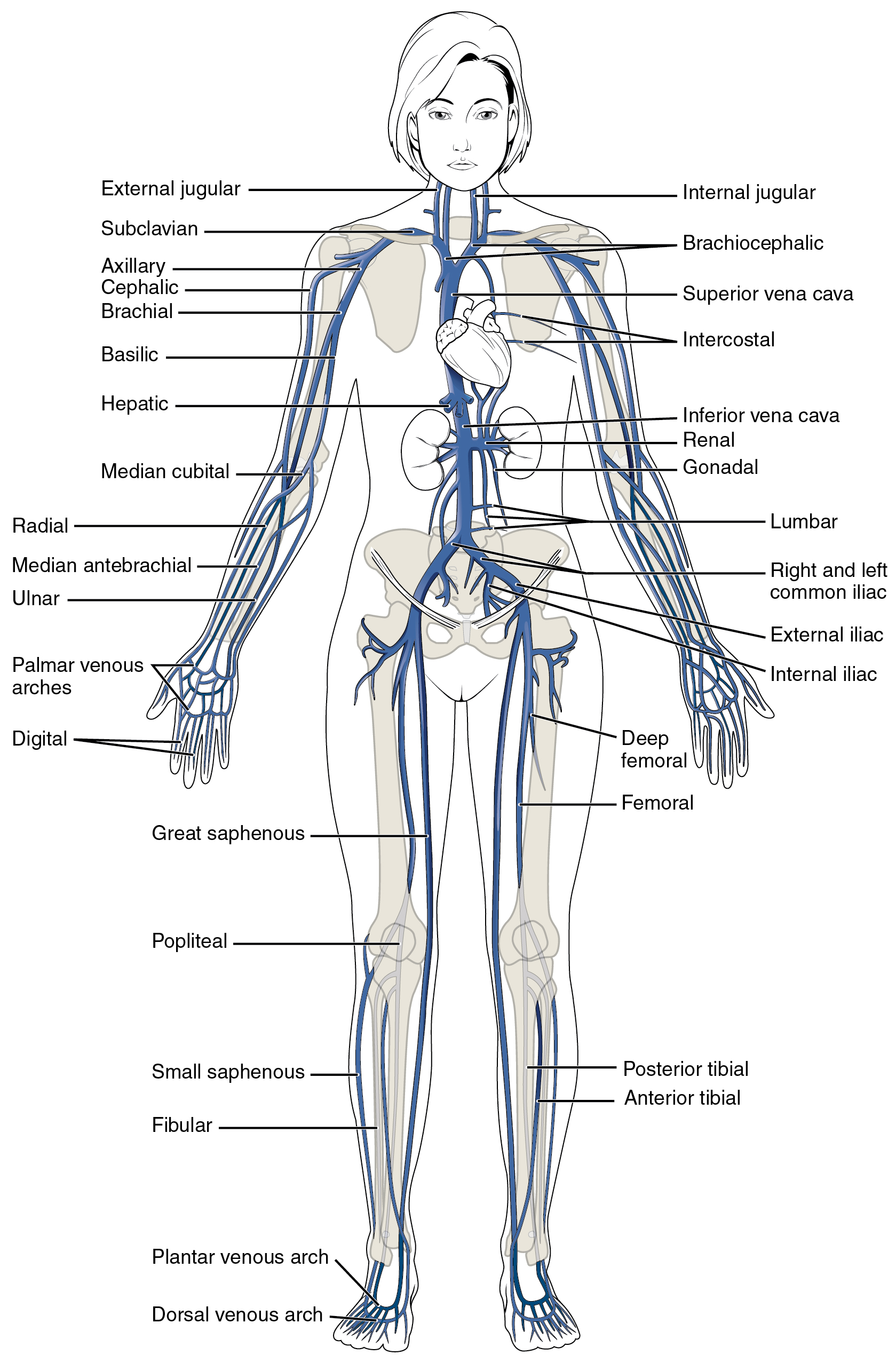
The venae comitantes of the brachial artery ie the deep veins of the arm or brachial veins are joined by the basilic vein above the lower border of the posterior wall of the axilla to form the axillary vein. It can anatomically be divided into the superficial veins and the deep veins.
 Upper Extremity Venous Thrombosis Thoracic Key
Anatomy physiology module provides a broad spectrum of adult male and female normal anatomy cases with varying body morphologies to maximize training efficacy.
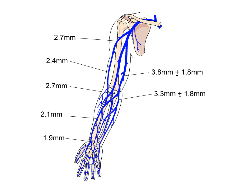
Venous anatomy arm. The venous system of the upper limb drains deoxygenated blood from the arm forearm and hand. The arteries deliver freshly oxygenated blood to muscles and bone. Forearm veins radial ulna still with the patient seated on the side of the bed follow the radial and ulnar veins to the wrist confirming compressibility and flow.
Within the venaecomitantes are both radian and ulnar veins with the ulnar veins typically existing as larger in size while the radial veins interact with the dorsal metacarpal veins. On the other hand the ulnar veins interact more with tributaries that deal with deep volar venous arches causing them to have more to do with the wrist area of a human being. At the antecubital fossa the brachial vein will divide into the radial ulnar veins.
Each individual hands on training case is accompanied by image window specific expert instruction and probe positioning guidance. There are three parts of the axillary vein the first distal part into which the cephalic vein enters at a point just superior to the pectoralis minor muscle and the second and third parts which give off branches corresponding to the tributaries off the axillary artery. The vessels of the arms are part of the circulatory system which provides nutrients to the tissues.
 Figure A1 A Central Venous Anatomy B Upper Extremity
 Figure 1 From Lower Extremity Venous Anatomy Semantic Scholar
 Anatomy Atlases Illustrated Encyclopedia Of Human Anatomic
 Arm Veins For Venipuncture Veins Dorsal Aspect Of The
 Block 4 Lsn 30 Arm Veins Diagram Quizlet
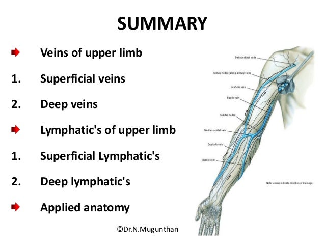 Venous Lymphatic Drainage Of Upper Limb Dr N Mugunthan
 Chapter 33 Venous And Intraosseous Access In Adults
 The Peripheral Veins Radiology Key
![]() Veins Of The Upper Limb Anatomy Kenhub
Veins Of The Upper Limb Anatomy Kenhub
 Vein Wikipedia
 How To Undertake Venepuncture To Obtain Venous Blood Samples
 Venous Cannulation Sites In The Arm Illustration Stock
 Picc Line Vein Anatomy Upper Limb Anatomy Anatomy Images
 23 Anatomy For Venipuncture Pocket Dentistry
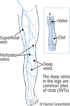 Deep Vein Thrombosis Blood Clots In Your Veins Harvard Health
 Cephalic Vein Wikipedia
 Clinical Education Intravenous Therapy Skills
 Forearm Veins Google Search In 2019 Arm Anatomy
Anatomy Atlases Anatomy Of First Aid A Case Study Approach

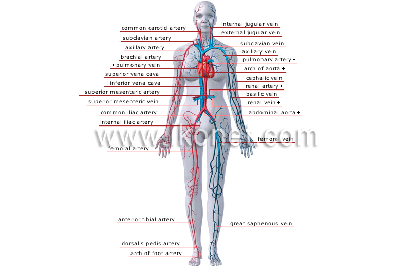
 20 5 Circulatory Pathways Anatomy And Physiology
 Venipuncture Module 1 Anatomy Of The Arm And Vein Location
 Upper Limb Veins 3d Anatomy Tutorial
 Ultrasonography
 How To Draw Blood Like A Pro Nurse Org
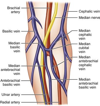
 Median Cubital Antebrachial Veins Locations Functions
 Upper Limb Anatomy
 Sonographic Evaluation Of Upper Extremity Deep Venous
 Arm Dvt Normal Ultrasoundpaedia
 Elbow Arm Anatomy

Belum ada Komentar untuk "Venous Anatomy Arm"
Posting Komentar