Anatomy Of The Female Pelvis
The female pelvic organs include the egg producing ovaries and the uterine tubes that carry the eggs into the uterus for potential fertilization by male sperm. The pelvis is composed of pairs of bones which are fused together so tightly that the joints are difficult to see.
 Female Pelvic Anatomy Medical Illustration Medivisuals
Anteroposterior compressions lateral compressions vertical shears combined fractures.

Anatomy of the female pelvis. The female urethra the female urethra runs from the internal urethral orifice of the urinary bladder anterior to. Also a couple of ligaments in the pelvis participate in forming the pelvis cavity. The female sacrum is wider shorter and less curved and the sacral promontory projects less into the pelvic cavity thus giving the female pelvic inlet pelvic brim a more rounded or oval shape compared to males.
The pelviss frame is made up of the bones of the pelvis which connect the axial skeleton to the femurs and therefore acts in weight bearing of the upper body. Female pelvis muscles puborectalis. The womans pelvis is adapted for child bearing and is a wider and flatter shape than the male pelvis.
The endometrium uterus ovaries cervix vagina and vulva. The female pelvic area contains a number of organs and structures. The book begins with a description of the functional anatomy of the pelvis how it responds to pregnancy childbirth.
Free shipping on qualifying offers. The female pelvic area contains a number of organs and structures. It is clear that the anatomy of pelvis is complex and consists of the several bones that are connected with mutual joints.
The lesser pelvic cavity of females is also wider and more shallow than the narrower deeper and tapering lesser pelvis of males. We will describe each of the bones in turn and their major landmarks. The pelvis is the lower portion of the trunk located between the abdomen and the lower limbs.
The iliococcygeus has thinner fibers and serves to lift the pelvic floor as well as. This muscle is responsible for holding in urine and feces. They also include the vagina which is the entryway to the uterus.
This is followed by a series of specific exercises. The endometrium uterus ovaries cervix vagina and vulva. This muscle makes up most of the levator ani muscles.
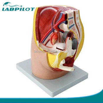 Median Sagittal Section Of Female Pelvic Model 3 Parts Human Female Pelvis Anatomy Model Buy Median Sagittal Section Of Female Pelvic Female
 Anatomy Of The Female Pelvis The Bmj
5 Anatomical Detail Of Female Pelvic Anatomy Adapted From
Department Of Urology At Miller School Of Medicine
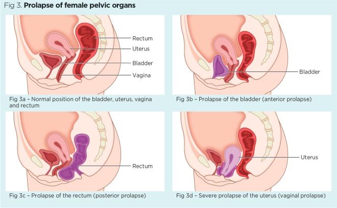 Female Pelvic Floor 1 Anatomy And Pathophysiology Nursing
 Vascular Anatomy Of The Female Pelvis
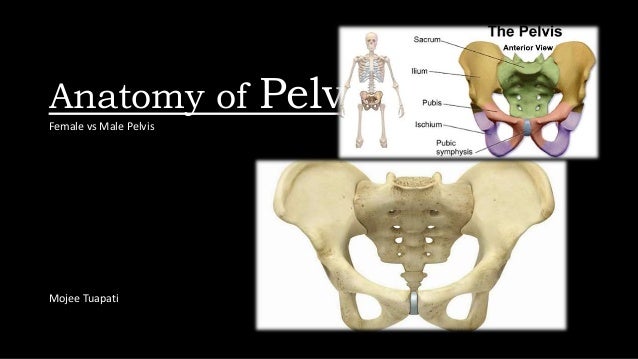 Pelvic Bones Anatomy Male Vs Female Pelvis
 Female Pelvis Anatomy Superficial View And Deep View
 Female Pelvis Model With Ligaments Vessels Nerves Pelvic Floor And Organs Life Size 6 Part
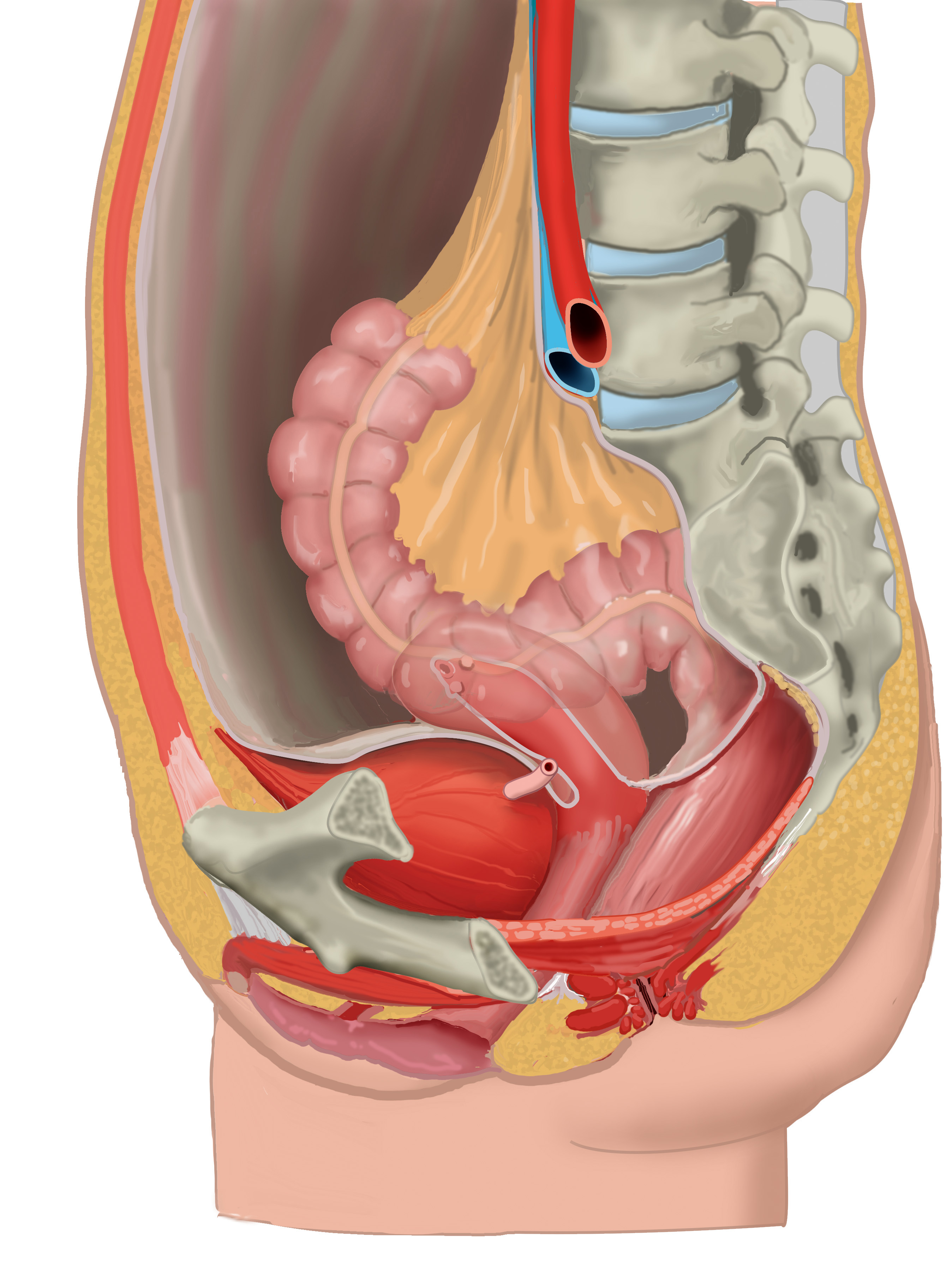 Sagittal Section Female Pelvis Anatomytool
 Female Pelvic Anatomy Medical Exhibit Medivisuals
 Anatomy Of The Female Pelvis Medical Illustration Human
 Anatomical Model Female Pelvis
 Female Pelvis With Detachable Pelvic Floor Muscles 12 Part
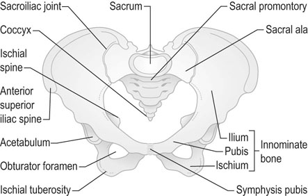 Anatomy Of The Female Pelvis Clinical Gate
 Female Pelvic Anatomy Repro With Otey At University Of
 Human Animal Anatomy And Physiology Diagrams Normal Anatomy
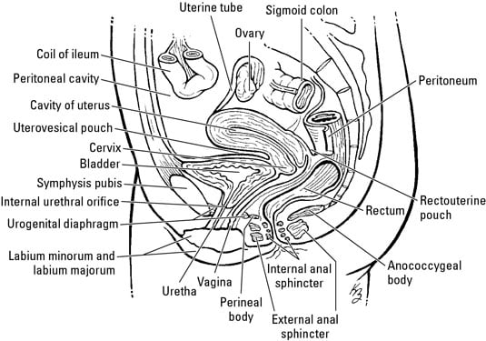 The Anatomy Of The Female Pelvis Dummies

 Blood Vessels Of The Female Pelvis Preview Human Anatomy Kenhub
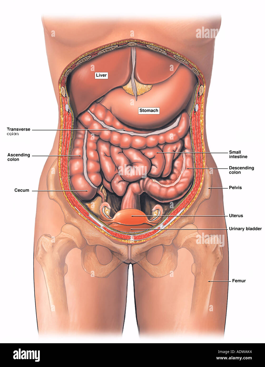 Anatomy Of The Female Abdomen And Pelvis Stock Photo
 Anatomy Pelvis Female Anatomy Of Pelvis In Woman Anatomy

Belum ada Komentar untuk "Anatomy Of The Female Pelvis"
Posting Komentar