Internal Ear Anatomy
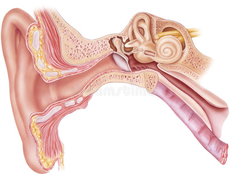
The chambers are full of fluid which vibrates when sound comes in and causes the small hairs which line the membrane to vibrate and send electrical impulses to the brain. The ear is made up of three parts.
 Anatomy Of Inner Ear By Dr Aditya Tiwari
The cochlea which is the hearing portion and the semicircular canals is the balance portion.

Internal ear anatomy. Middle ear tympanic cavity anatomy duration. External ear auricle see the following image external ear anatomy. The inner ear is the innermost part of the ear which consist of the cochlea the balance mechanism the vestibular and the auditory nerve.
In vertebrates the inner ear is mainly responsible for sound detection and balance. The anatomy of the ear is composed of the following parts. They send information on balance and head position to the brain.
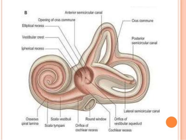
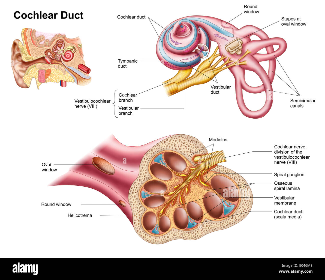
The modiolus is perforated spirally at its base in the internal acoustic meatus by the fibres of the cochlear nerve. The fluid filled semicircular canals labyrinth attach to the cochlea and nerves in the inner ear. Ears also help to maintain balance.
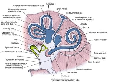
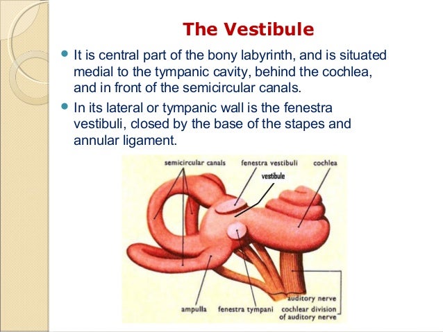
In mammals it consists of the bony labyrinth a hollow cavity in the temporal bone of the skull with a system of passages comprising two main functional parts. Pain in the ear can have many causes. It lies between the middle ear and the internal acoustic meatus which lie laterally and medially respectively.
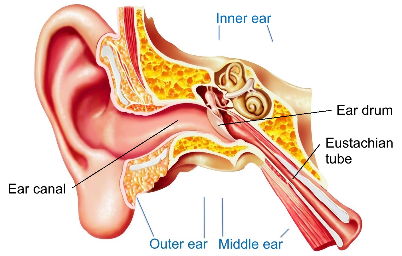
Read more in this article about the inner ears anatomy how the inner ear functions and the parts of the inner ear. The eustachian auditory tube drains fluid from the middle ear into the throat pharynx behind the nose. The inner ear internal ear auris interna is the innermost part of the vertebrate ear.
The base of modiolus is located at the fundus of the internal acoustic meatus and apex points in the direction of the middle ear. Sam webster 37718 views. The outer middle and inner ear.
Ear anatomy inner ear. The inner ear is located within the petrous part of the temporal bone. The inner ear has two main components the bony labyrinth and membranous labyrinth.
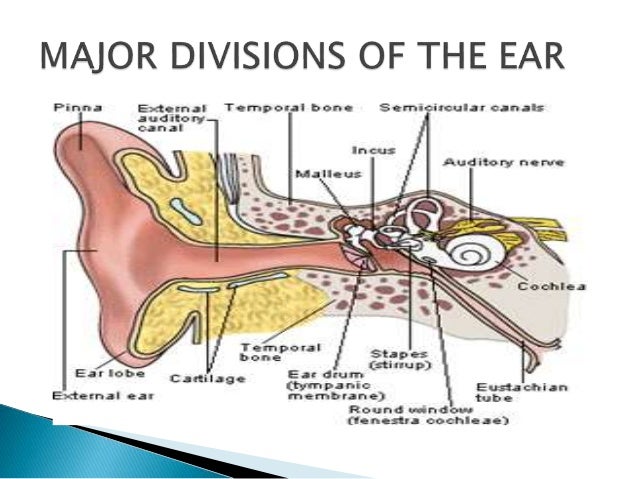
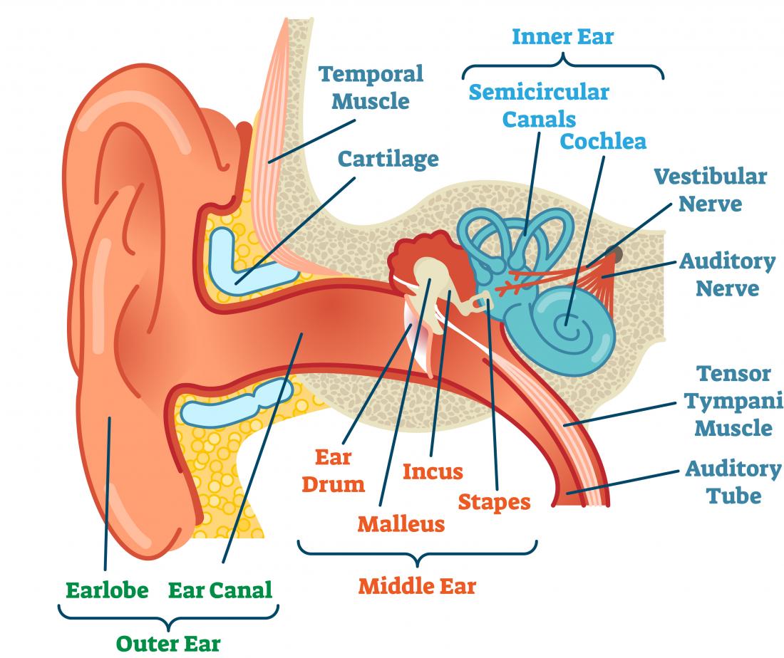
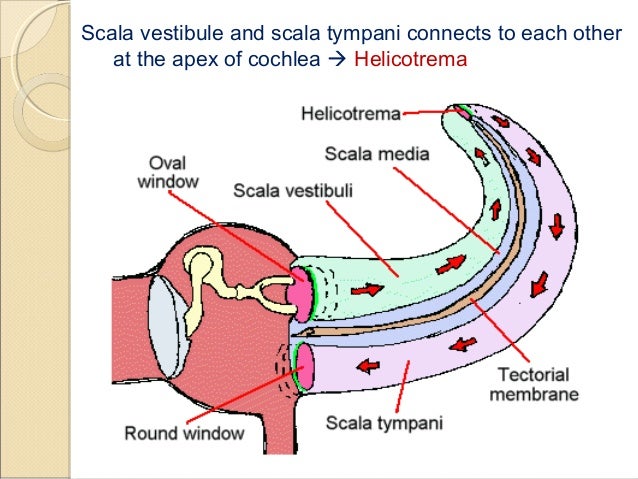
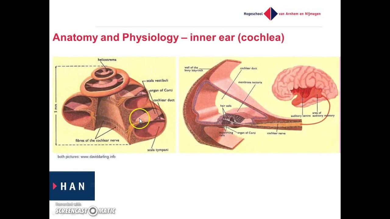
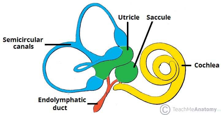
The cochlea is shaped like a snail and is divided into two chambers by a membrane. Inner ear also called labyrinth of the ear part of the ear that contains organs of the senses of hearing and equilibrium. Semicircular canals vestibule cochlea see the image below cross section of the middle and inner ear.
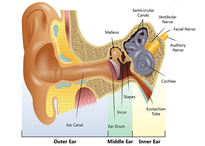
The apex of the modiolus is overlaid by the apical turn of the cochlear canal. All three parts of the ear are important for detecting sound by working together to move sound from the outer part through the middle and into the inner part of the ear. The bony labyrinth a cavity in the temporal bone is divided into three sections.
The vestibule the semicircular canals and the cochlea. Malleus incus and stapes see the image below inner ear labyrinthine.
 Total Ear Canal Ablation Teca In Dogs
 Reference Chart Anatomy Of The Inner Ear Biologyproducts Com
Picture Of Ear Anatomy Picture Image On Rxlist Com
 Ear Anatomy Cross Vector Photo Free Trial Bigstock
 Ear Anatomy Overview Embryology Gross Anatomy
 Cartoon Of Human Internal Ear Anatomy
 Ears And Hearing How Do They Work
 Inner Ear Anatomy
 Anatomy Of The Inner Ear And Facial Nerve By Annie Campbel
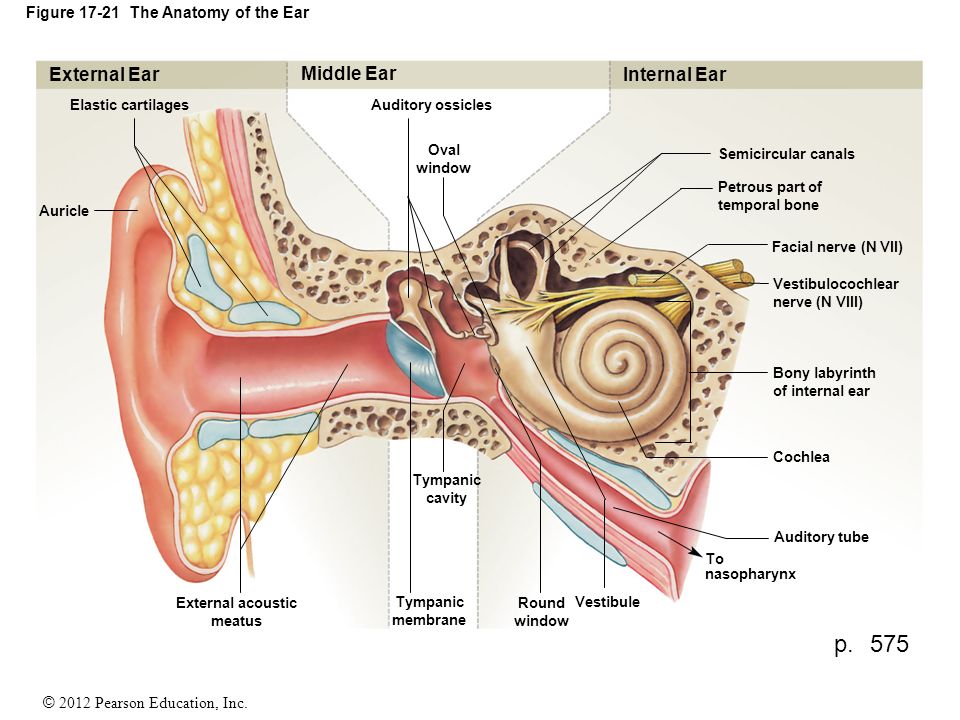 2012 Pearson Education Inc Figure The Anatomy Of The Ear
 Anatomy Of Inner Ear
 Types Of Hearing Impairment University Of Iowa Hospitals
 Anatomy And Physiology Of The Inner Ear Youtube
 Ear Wikipedia
 Internal Ear Stock Photos Internal Ear Stock Images Alamy
 Parts Of Inner Ear Anatomy Diagram Reading Industrial
 The Inner Ear Bony Labyrinth Membranous Labryinth
 Understanding How The Ear Works Hearing Link
 Introduction To The Anatomy And Physiology Of The Auditory
 Anatomy Of Inner Ear
 Hearing And Hair Cells
 Ear Infection Middle Ear Symptoms Treatment Southern
 Ear Disorders Problems And Treatment Ent Florida
 Anatomy Of The Inner Ear
 Anatomy Ear Outer Middle Inner
 Ear Anatomy Stock Illustration Illustration Of Quiet


Belum ada Komentar untuk "Internal Ear Anatomy"
Posting Komentar