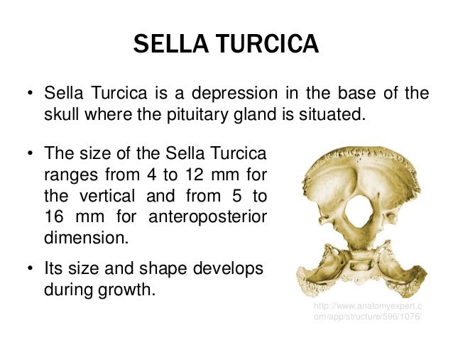
Sella Turcica Anatomy
In this review after a brief explanation of the anatomical and endocrinological features of the sella turcica had been given a historical perspective of sella turcica nomenclature was presented for the first time. Medical definition of sella turcica.
 Dorsum Sellae And Sella Turcica Google Search Portal
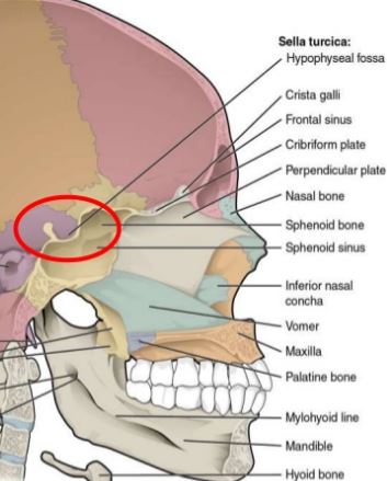
The sella turcicas most inferior portion is known as the hypophyseal fossa the seat of the saddle and contains the pituitary gland hypophysis.

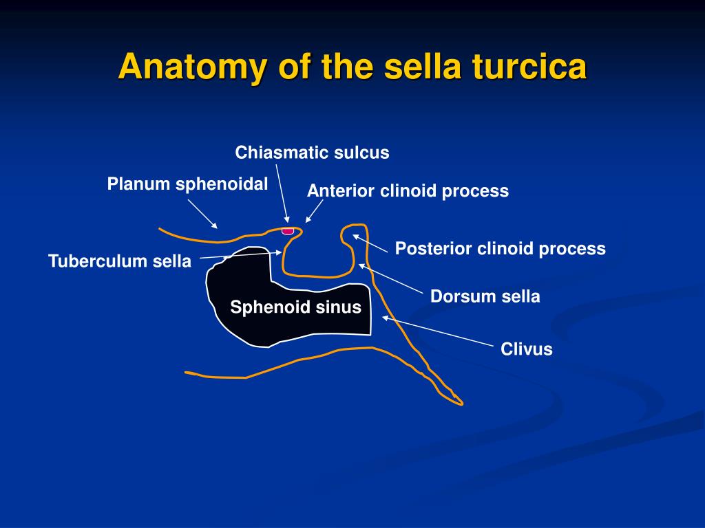
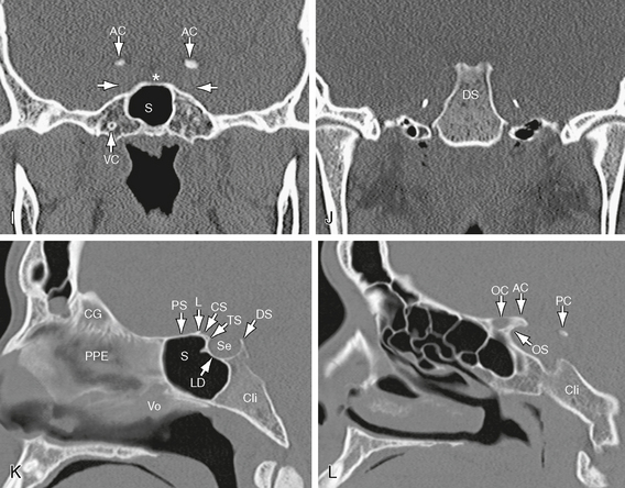
Sella turcica anatomy. The sphenoid bone sph forms the bony floor of the sella turcica. The sella turcica is structurally a cup shaped recession which is located in the central basisphenoid bone that encloses the pituitary gland along with inferior portion of the infundibular stalk. Coronal cryomicrotome section through the sella turcica.
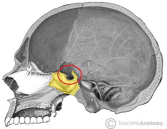
The sella turcica is located deep within the cranium but can be demonstrated on a number of projections used in skull radiography. Location of sella turcica in skull. Gross anatomy the anterior inferior and posterior walls are bony while the lateral walls and roof a.
There are two general types of pituitary tumourshormone secreting and nonsecreting. Resident doctor jpntc aiims. Other articles where sella turcica is discussed.
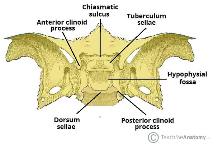
The sellar floor is anteriorly constant with the tuberculum sellae and the dorsum sellae posteriorly. Figure 14 4 gross anatomy of the sella and pituitary gland. The pituitary hypophyseal fossa or sella turcica is a midline dural lined structure in the sphenoid bone which houses the pituitary gland.
Sella turcica simplified anatomy presented by dr. A depression in the middle line of the upper surface of the sphenoid bone in which the pituitary gland is lodged. Afshan jabeen former jr.
Providing the proper place to hold and support the pituitary gland. 3 clinical anatomy of sella turcica. This picture of the skull with temporal and parietal bones removed shows the location of the sella in red.
Cause of enlargement of the sella turcica the bone cavity in the head in which the pituitary gland is located. There are five types of hormone secreting pituitary tumours named according to the cells that produce the particular hormone. The name sella turcica is one of the most commonly used terms in everyday endocrine practice.
It belongs to the middle cranial fossa. Anatomy of the sella turcica. The sella turcica is located in the sphenoid bone behind the chiasmatic groove and the tuberculum sellae.
The purplish pituitary gland unlabeled rests on the floor of the sella between the paired cavernous segments of the internal carotid arteries ic. Functions of sella turcica. Any anomaly or pathological condition in the pituitary gland can apparent from a distorted form of sella turcica to a disorder in the malfunctioning of the secretion of the hormones secreted by the pituitary which includes.
 Incidental Finding Of An Enlarged Sella Turcica On A Lateral
 Sella Turcica Sphenoid Bone Skull Anatomy Anatomy
 The Morphology Of Sella Turcica In Individuals With

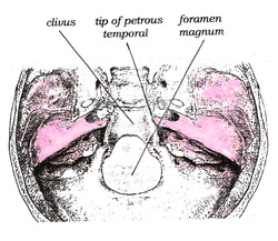 Clivus Anatomy Wikipedia
 Sella Turcica Rad32
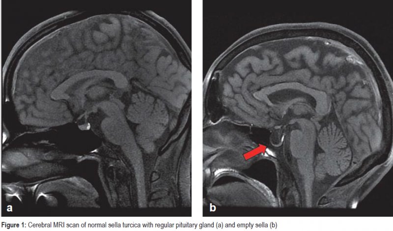
 Empty Sella Syndrome Information Mount Sinai New York
 Sella Turcica Archives Maryann Reynolds Ms Lmt Bctmb
 Turcica Ellsworth Sessions
 Middle Cranial Fossa Boundaries Contents Teachmeanatomy
 Cerebral Aneurysms With Intrasellar Extension A Systematic
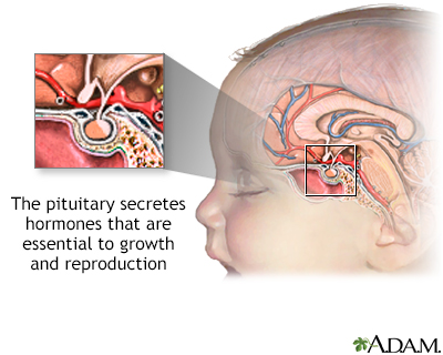
:max_bytes(150000):strip_icc()/pituitary_gland-5a0c7e374e4f7d0036270998.jpg) Pituitary Gland Function And Hormone Production
Pituitary Gland Function And Hormone Production
 Ppt Sella Turcica And Parasellar Region Powerpoint
 Sella Turcica Wikipedia
 The Radiology Assistant Sella Turcica And Parasellar Region
 The Radiology Assistant Sella Turcica And Parasellar Region
Sella Turcica
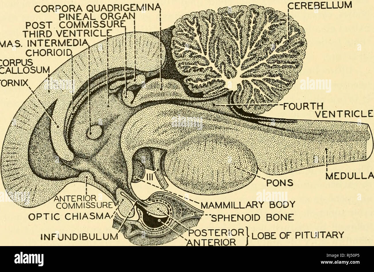 Chordate Anatomy Chordata Anatomy Comparative 534
 Turcica Ellsworth Sessions
Anatomy And Imaging Of The Normal Sella Turcica And
 Sella Turcica And Pituitary Gland Radiology Key
 Sphenoid Bone Location Structure Function Teachmeanatomy
 Sella Turcica Wikiwand
 Empty Sella Radiology Reference Article Radiopaedia Org
Belum ada Komentar untuk "Sella Turcica Anatomy"
Posting Komentar