Shoulder Mri Anatomy
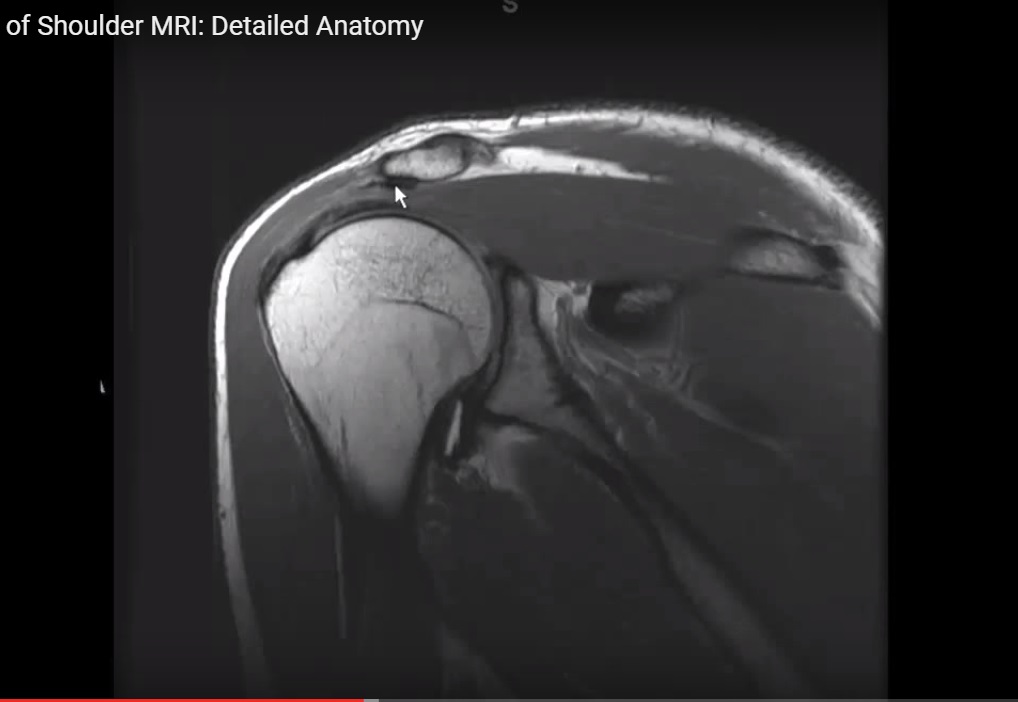
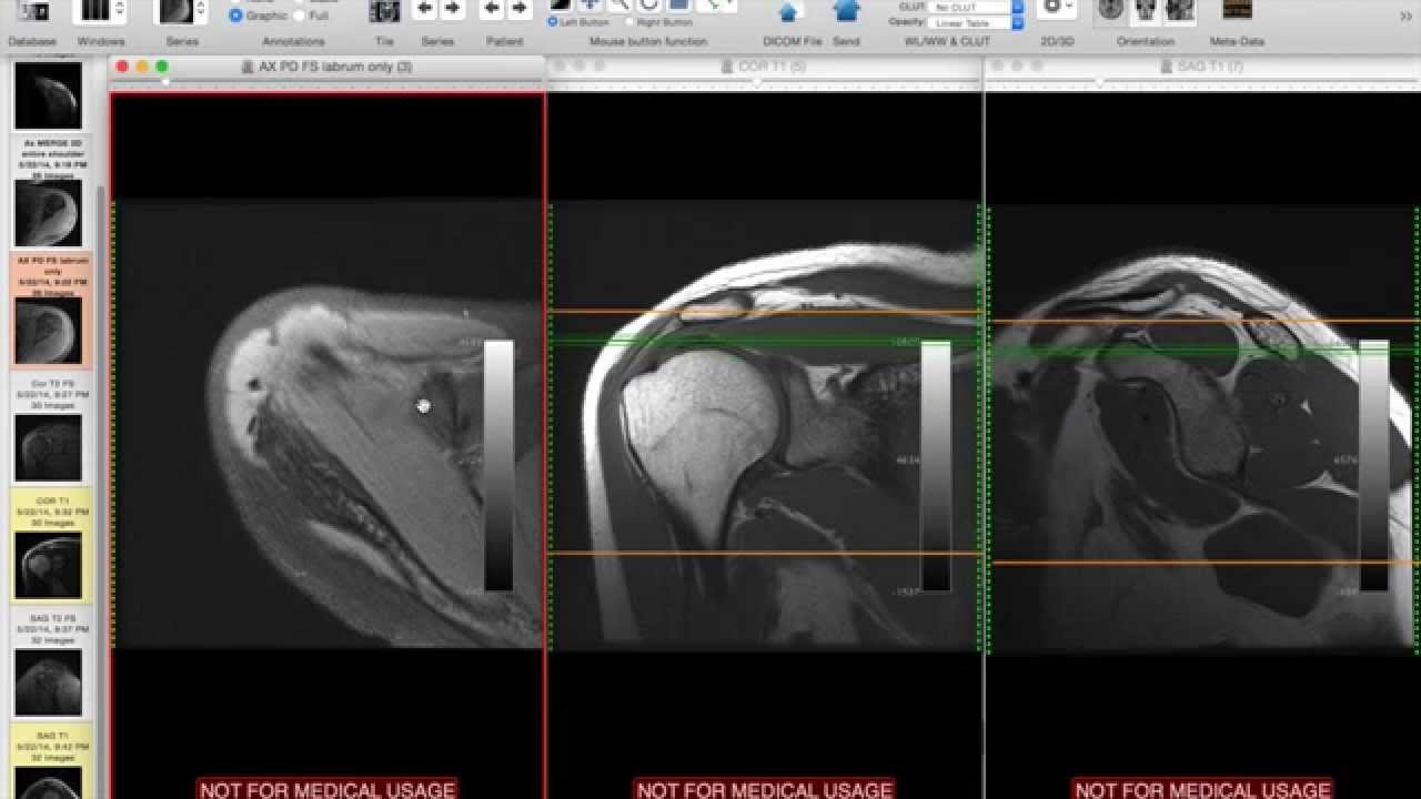
Mr is the best imaging modality to examen patients with shoulder pain and instability. Use the mouse scroll wheel to move the images up and down alternatively use the tiny arrows on both side of the image to move the images on both side of the image to move the images.
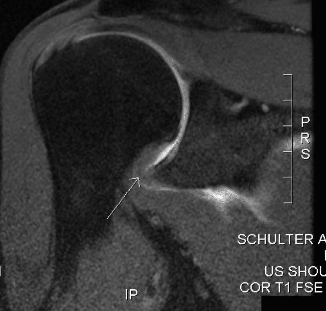
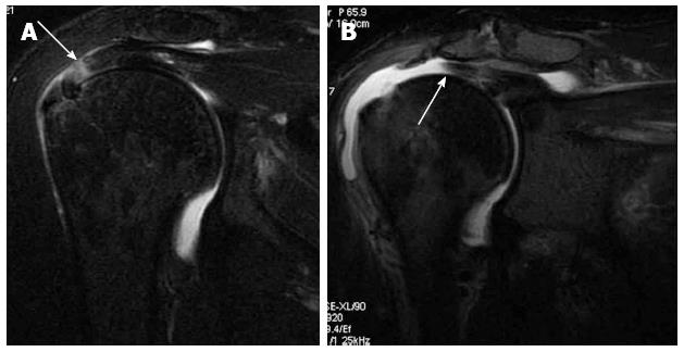
T2 star gradient recall echo images are employed in the assessment of the labrum and for detection of substances that produce susceptibility effects such as calcium hydroxyapatite or loose surgical hardware.

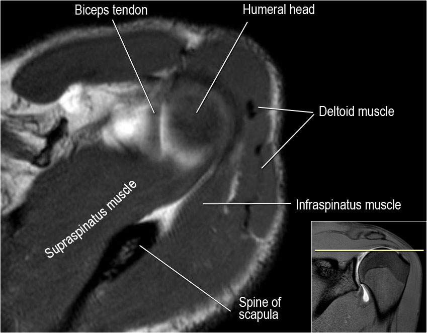
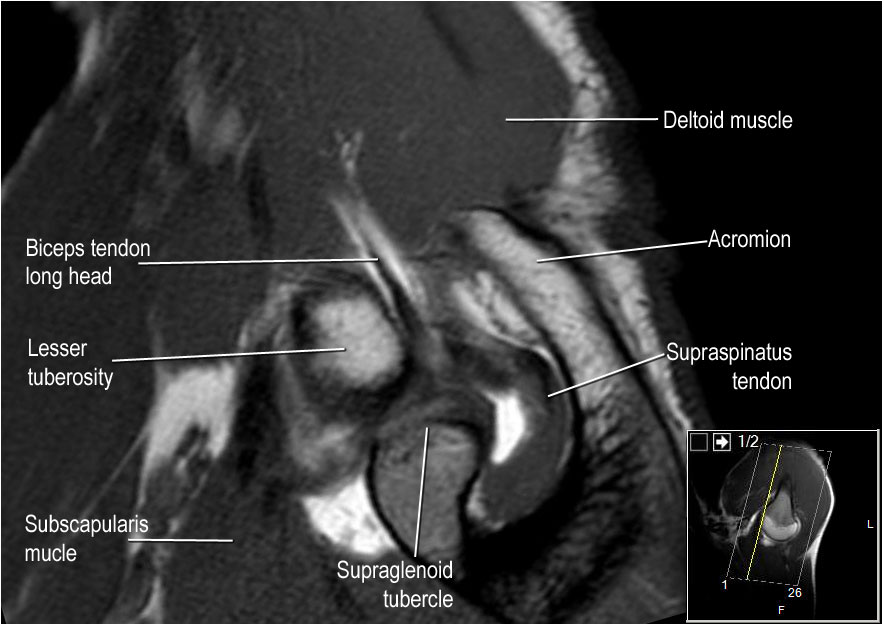
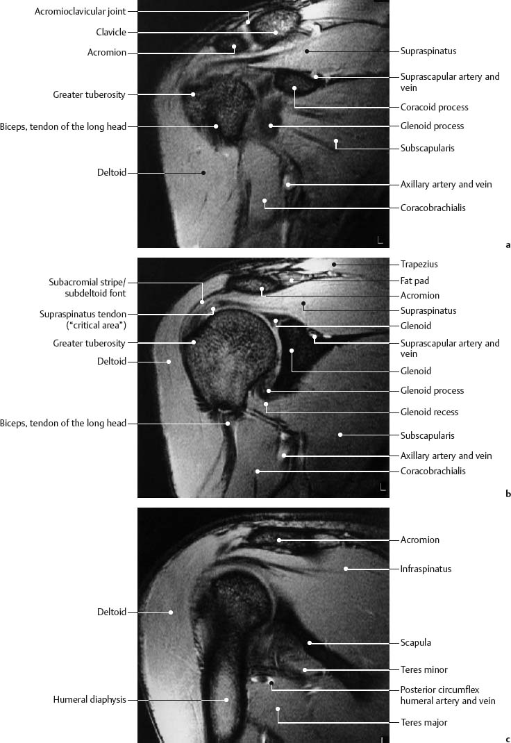
Shoulder mri anatomy. An mri of the shoulder of a healthy subject was performed in the 3 planes of space coronal axial sagittal commonly used in osteoarticular imagery with two weightings most commonly used to explore the musculo skeletal pathology of the shoulder. About anatomy mri magnetic resonance imaging is particularly well suited for the medical evaluation of the musculoskeletal msk system including the knee shoulder ankle wrist and elbow. Review of basic normal imaging anatomy of the shoulder on non contrast mri highlighting the important structures to analyze during readout.
From the chief of msk radiology stanford university. Spin echo t1 and proton density with fat saturation sequences. Knee shoulder shoulder arthrogram ankle elbow wrist hip.
Atlas of shoulder mri anatomy. The glenohumearal joint has a greater range of motion than any other joint in the body. These bones combine to make three sub joints.
Injuries such as anterior cruciate ligament meniscus and rotator cuff tears are all easily diagnosed when there is a firm understanding and knowledge of human anatomy. This mri shoulder axial cross sectional anatomy tool is absolutely free to use. Stanford bone tumor bayesian network issssr msk lectures for residents ocad msk cases from around the world stanford msk mri atlas has served almost 800000 pages to users in over 100 countries.
Click on a link to get t1 axial view t2 fatsat axial view t1 coronal view t2 fatsat coronal view t2 fatsat sagittal view. This webpage presents the anatomical structures found on shoulder mri. Use the mouse to scroll or the arrows.
Shoulder anatomy is formed by the union of three major bones including the humerus scapula and clavicle. The small size of the glenoid fossa and the relative laxity of the joint capsule renders the joint relatively unstable and prone to subluxation and dislocation. Mri shoulder protocols typically involve fat saturated proton density images that are sensitive to internal derangement.
 Teaching Files University Of North Dakota

 Shoulder Impingement 3 Keys To Assessment And Treatment
 The Radiology Assistant Shoulder Mr Anatomy

 The Radiology Assistant Shoulder Mr Anatomy
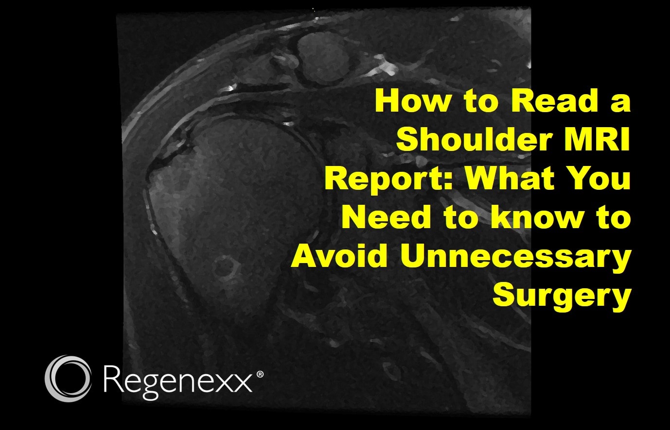 How To Read A Shoulder Mri Report Regenexx
 The Radiology Assistant Shoulder Mr Anatomy
 Improve Msk Imaging Outcomes Imaging Technology News
Shoulder Mri Radiographical And Illustrated Anatomical Atlas
 Normal Mri Anatomy Of The Musculoskeletal System Radiology Key
 Mri Of Shoulder Anatomy
 Shoulder Anatomy Mri Shoulder Axial Anatomy Free Cross
 Radiology Anatomy Images Sagittal Anatomy Of Shoulder Mri
 Shoulder Radiology Key
 Normal And Variant Anatomy Of The Shoulder On Mri
 Adhesive Capsulitis Frozen Shoulder Management
 Shoulder Anatomy And Normal Variants
 Shoulder Mri Radiographical And Illustrated Anatomical Atlas
 Systematic Interpretation Of Shoulder Mri How I Do It
 Ppt Mri Anatomy Of The Shoulder Powerpoint Presentation
 Rotator Cuff Disorders How To Write A Surgically Relevant
 Shoulder Anatomy Mri Shoulder Axial Anatomy Free Cross
 Shoulder Mri Radiographical And Illustrated Anatomical Atlas
Belum ada Komentar untuk "Shoulder Mri Anatomy"
Posting Komentar