Calf Anatomy
Started in 1995 this collection now contains 6731 interlinked topic pages divided into a tree of 31 specialty books and 732 chapters. The gastrocnemius has two heads.

Sura is the back portion of the lower leg in human anatomy.
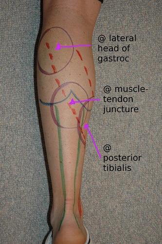
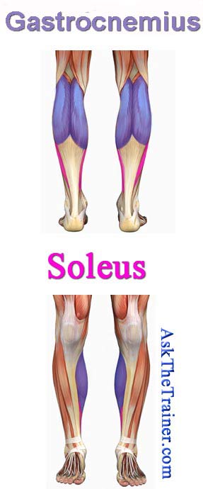
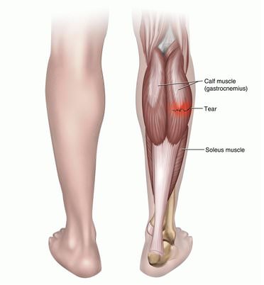
Calf anatomy. The calf anatomy includes the gastrocnemius aka gastroc and the soleus. The gastrocnemius has two parts or heads which together create its diamond shape. The calf muscle on the back of the lower leg is actually made up of two muscles.
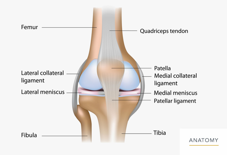
The ankle is made off the tibia and fibula of the leg as well as the talus of the foot. The medial and the lateral. Anatomy of the calf muscles.
The ankle is a joint that connects the lower leg to the foot. Discover more information about the calf anatomy by clicking the links throughout the page. Each of these muscles is a discrete organ constructed of skeletal muscle tissue blood vessels tendons and nerves.
It attaches to the heel with the achilles tendon and originates behind the knee on the femur crossing two joints. Anatomy of the calf muscle. Get ready to learn about both of these muscles their locations and their functional anatomy.
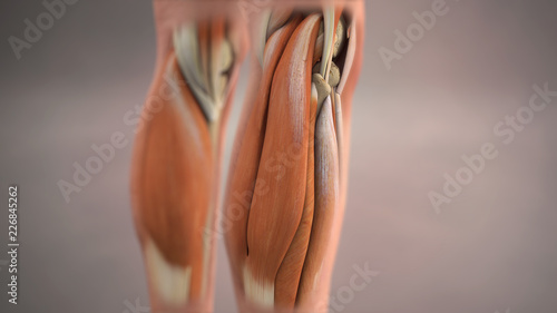
The gastrocnemius is the calf muscle that is visible from the outside of the body. The muscles within the calf correspond to the posterior compartment of the leg. The gastrocnemius is the larger calf muscle forming the bulge visible beneath the skin.
This page provides an overview of the calf muscle group. People tend to be tight in the back of the body splayed open if you will. Attached to the bones of the skeletal system are about 700 named muscles that make up roughly half of a persons body weight.
Its main function is to allow for plantar flexion and dorsiflexion of the foot. From top to bottom the neck and lower back muscles along with the gluteus maximus hamstrings calves and achilles tendon are all tight in relationship to their opposite muscles at the front of the body. The soleus is a smaller flat muscle that lies underneath the gastrocnemius muscle.
The muscular system is responsible for the movement of the human body. The two largest muscles within this compartment are known together as the calf muscle and attach to the heel via the achilles tendon.
 Calf Leg Wikipedia
 Calf Anatomy
 Calf Rom Stretching
 Anatomy Of The Calf Lower Leg Muscles Posterior Diagram


 The Importance Of Calf Strength And The Best Calf Strength
 Anatomy Of The Calf Muscles Brevis Extensor Digitorum Longus Gastrocnemius
 Best Calf Exercises Exercise Tips To Build Strong Calve

 High Calf And Low Calf The Stephane Andre


 How To Stretch Calf Muscles And The Big Mistake To Avoid
 Knee Calf Orthopedic Specialist Of Northern California
 Muscle Anatomy Skeletal Muscles Groin Muscles Calf Muscles
Achilles Tendonitis Warning Signs Of Achilles Tendon Injury
 Calf Muscles Anatomy Medical Illustration Buy This Stock


 Pin On Fitness
 Calves Premium Assignment Examples Proko
 Calf Anatomy All About The Calf Muscles
 Muscles Of The Lower Leg And Foot Human Anatomy And
 Anatomy Calf Muscles Diagram Quizlet

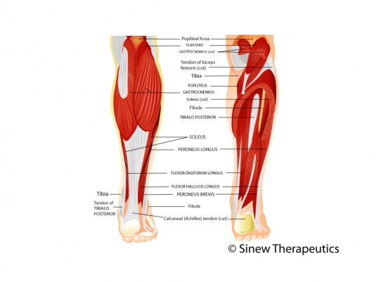 Calf Injuries Calf Pain Information Sinew Therapeutics
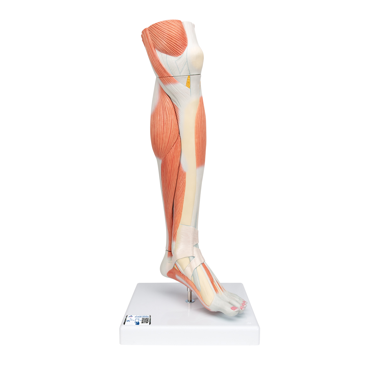 Anatomical Teaching Models Plastic Human Muscle Models
 How To Draw The Calf Proko
 Calf Strain Local Physio
Belum ada Komentar untuk "Calf Anatomy"
Posting Komentar