Pericardial Anatomy
The right phrenic nerve passes to the right of the pericardium. Vagus nerve cn x where the function is uncertain.
![]() Pericardium Anatomy Of Fibrous And Serous Layers Kenhub
Pericardium Anatomy Of Fibrous And Serous Layers Kenhub
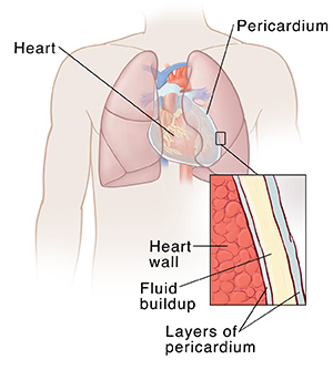
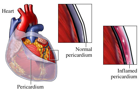
The pericardium is a fibroserous fluid filled sack that surrounds the muscular body of the heart and the roots of the great vessels.
Pericardial anatomy. Pleural and pericardial effusions. Pericardial anatomy and function the pericardium is a fibrous sac that surrounds the heart. Pericardium is the fluid filled sac that surrounds the heart and the proximal ends of the aorta venae cavae and the pulmonary artery.
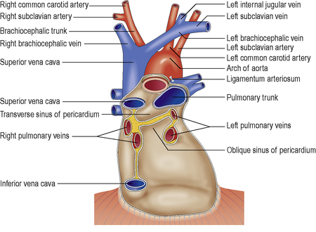
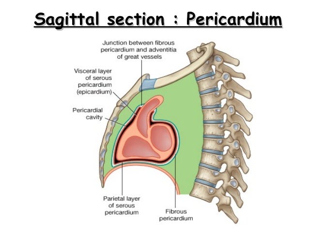
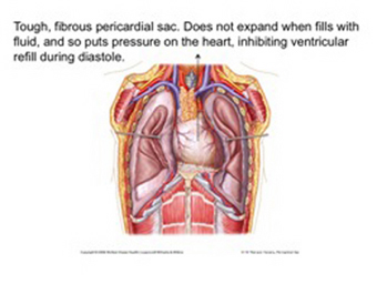
The heart and pericardium are situated behind the sternum breastbone in a position in the middle of the chest cavity known as the mediastinum. The serous pericardium fig. The pericardium is a conical flask like fibroserous sac which contains the heart and the roots of the great vessels and defines the middle mediastinum.
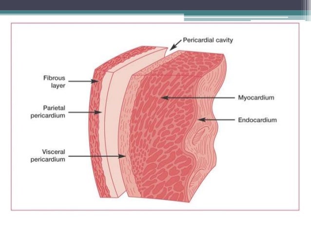
It consists of two layers. The normal pericardium is a fibroelastic sac surrounding the heart. The potential space between the parietal and visceral layers contains a thin film of fluid and is known as the pericardial cavity.
The sympathetic trunk which provides postganglionic vasomotor fibers. Anatomical relationships surrounds heart and bases of pulmonary artery and aorta. The outer sac is the fibrous pe.
This article will give an outline of its functions structure innervation and its clinical significance. 23 1 is a closed sac the parietal layer of which lines the inner surface of the fibrous pericardium and is reflected onto the heart as the visceral layer or epicardium. When it comes to innervation the pericardium has three main sources.
Phrenic nerves c3 c5 provide mostly somatic afferent pain temperature sensory. The normal pericardium is a fibroelastic sac surrounding the heart. Gross anatomy the pericardium is made of two sacs in one.
Deep to sternum and anterior chest wall. The pericardium is a dual layered structure enveloping. The visceral pericardium and the parietal pericardium.
Pericardial arteries supply blood. The left phrenic nerve passes over the pericardium of the left ventricle.
 7 Pericardial Structure And Function Thoracic Key
 1 Pericardium
 Pericardium Definition Function Layers Human Anatomy Kenhub
 Heart Anatomy Size Location Coverings And Layers
 Understanding Pericardial Effusion
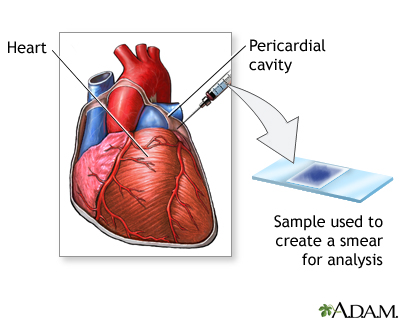 Pericardial Fluid Culture Medlineplus Medical Encyclopedia
 Pericardial Sac And Pericardial Fluid Anatomy
 Anatomy Block 2 Pericardium Heart Flashcards Quizlet
 Assessment Of Pericardial Effusion Differential Diagnosis
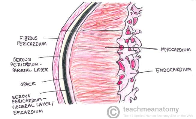 The Pericardium Teachmeanatomy
 Anatomy Of Pericardium
 Clinical Cases Pericardial Effusion
 Pericarditis Cormedicalgroup Com

 Pericardium Hea
 What Is The Structure Function And Location Of The Pleura

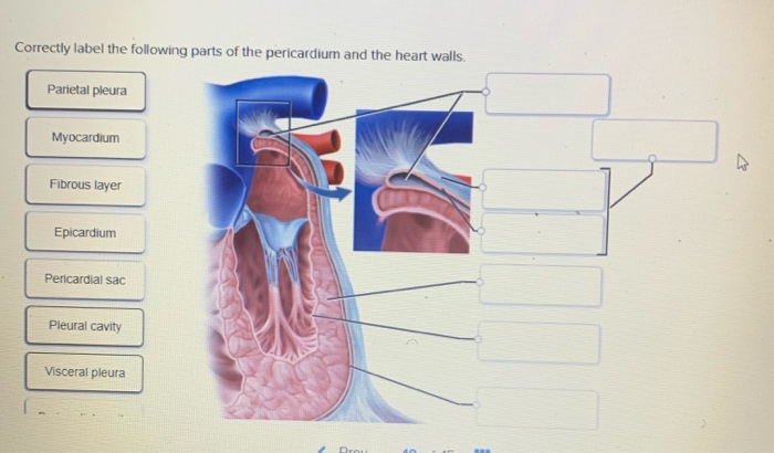 Solved Correctly Label The Following Parts Of The Pericar
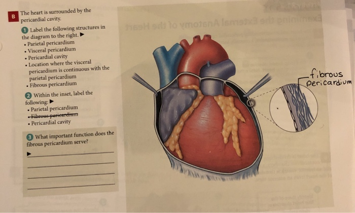
Solved The Heart Is Surrounded By The Pericardial Cavity
 Pericardial Cavity An Overview Sciencedirect Topics
 Pericardium Wikipedia
 Pericardial Pathology And Clinical Test 2 Flashcards Quizlet

 Pericardium Anatomy Britannica
 Pericardial Sac Picture Image On Medicinenet Com
 Layers Of The Heart Muscle And Pericardium The Section Of
Belum ada Komentar untuk "Pericardial Anatomy"
Posting Komentar