Ankle Mri Anatomy
Anterior posterior medial and lateral. Screen on fatsat images for bone marrow edema.

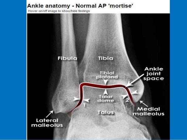
The ankle joint also known as talocrural joint is an example of a synovial joint and is formed by the bones tendons and ligaments found in the leg and the foot 1 2.
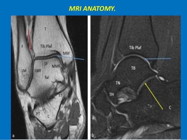
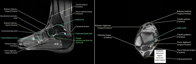
Ankle mri anatomy. It carries the weight of the body and can undergo a myriad of pathology most commonly traumatic injuries of the medial and lateral malleoli. There are several bones that make up the ankle. Mri anatomy of the ankle tendons and ligaments normal mri tendon anatomy tendons around the ankle are divided into four groups.
Ankle mri examination systematic approach. Mr imaging of the ankle and foot introduction. Once you have studied the bones scan the joints for effusion.
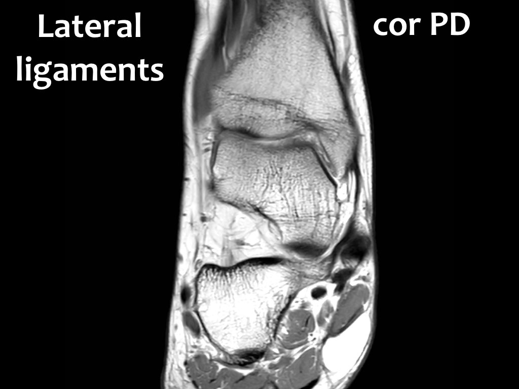
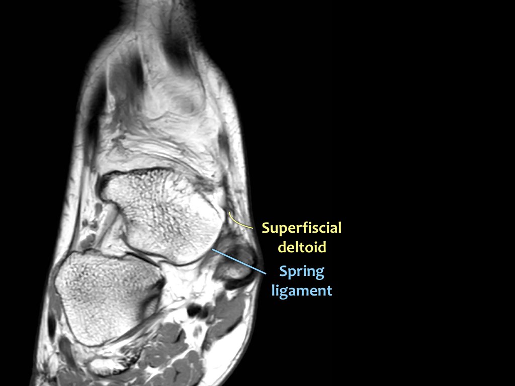
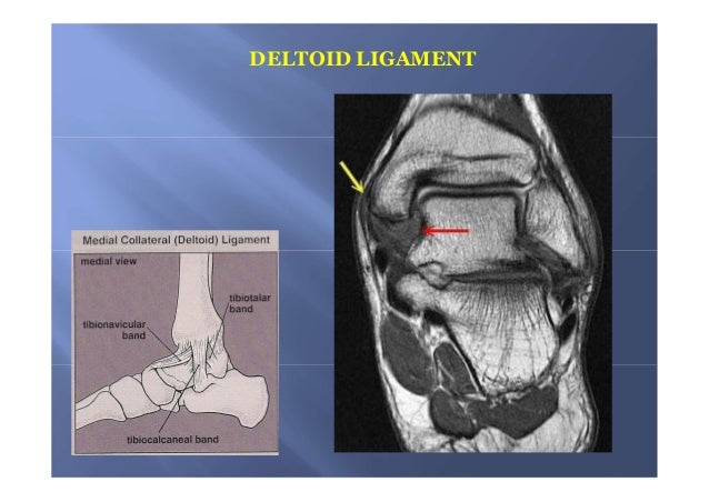
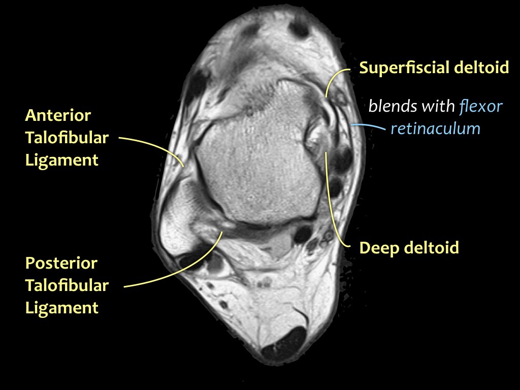
Mri of the ankle. Mri of the ankle and feet. Three ligamentous groups support the ankle joint.
About anatomy mri magnetic resonance imaging is particularly well suited for the medical evaluation of the musculoskeletal msk system including the knee shoulder ankle wrist and elbow. This webpage presents the anatomical structures found on ankle mri. Scroll through the image stack for the.
Start your exam with fatsat images of the bones to screen for edema. The tibia the fibula the talus and the calcaneus. The ankle joint is comprised of the tibia fibula and talus as well as the supporting ligaments muscles and neurovascular bundles.
Knee shoulder shoulder arthrogram ankle elbow. The past 15 years have witnessed an explosion of information regarding the role. The tibia extends inferiorly to articulate with.
Injuries such as anterior cruciate ligament meniscus and rotator cuff tears are all easily diagnosed when there is a firm understanding and knowledge of human anatomy. It is also a fundamental communication tool to teach patients anatomy and pathology. Use the mouse to scroll or the arrows.
This module is a comprehensive and affordable learning tool for medical students and residents and especially for physicians anatomists rheumatologists orthopaedic surgeons and radiologists. Click on a link to get sagittal view t1 axial view t2fatsat coronal view t2fatsat sagittal view t2fatsat. Routine ankle mr imaging is performed in the axial coronal.

 Presentation1 Pptx Ankle Joint
 Ankle Mri Anatomy Hnchawaii Org Mri Brain


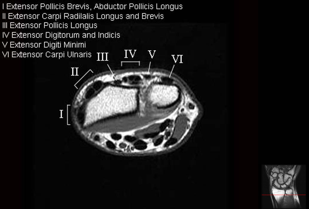 Mri Wrist Anatomy
 Ankle Mri
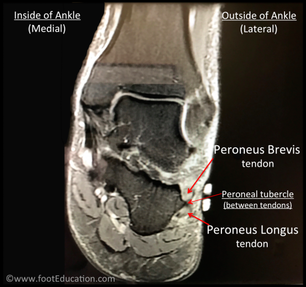 Peroneal Tendonitis Footeducation
 Mri Anatomy Of Ankle Radiology Case Radiopaedia Org


 Presentation1 Pptx Ankle Joint
 Mri Ankle Google Search Ankle Joint Ankle Anatomy Images
 The Radiology Assistant Ankle Mri Examination
 Ankle Mri Radiology Key
 The Radiology Assistant Ankle Mri Examination
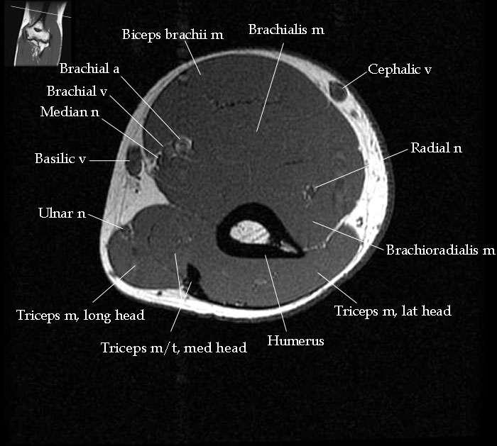
Mri Elbow Anatomy
 Mri Sliders Mri Anatomic Imaging Of The Ankle 1 Mr Tip Com
 The Radiology Assistant Ankle Mri Examination
 Mri Anatomy Of Ankle
 Ecr 2015 C 1824 Osteochondral Lesions Of The Talus A
 Update On Diagnosis And Management Of Cuboid Fractures
 Mri Anatomy Of Ankle
 Mri Ankle Unidad Especializada En Ortopedia Y Traumatologia

 Mri Anatomy Of Ankle Ligaments Deltoid Ligament
 The Radiology Assistant Ankle Mri Examination
 Anatomy Of The Foot And Ankle Mri

Belum ada Komentar untuk "Ankle Mri Anatomy"
Posting Komentar