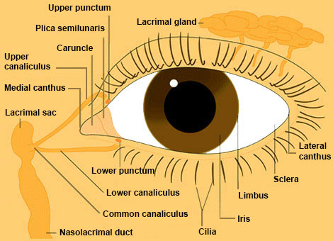
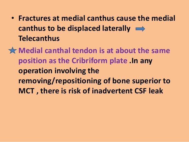
Medial Canthus Anatomy
Touching the lateral canthus of the eye evaluates the maxillary branch. It is 3 mm lower in asians.
 The Transnasal Bilobed Flap For Medial Canthal Reconstruction
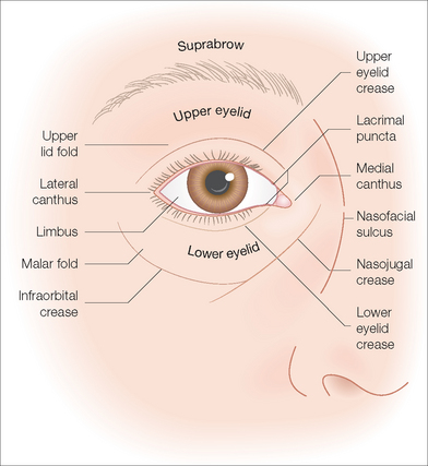
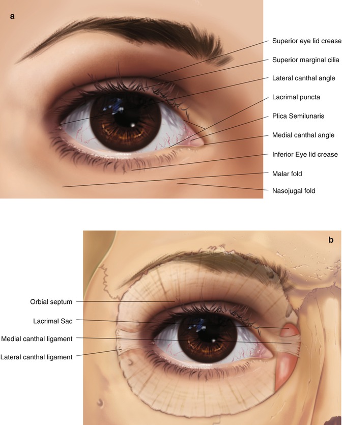
The one on the inner aspect is called the medial canthus while that at the outer aspect is called the lateral canthus.
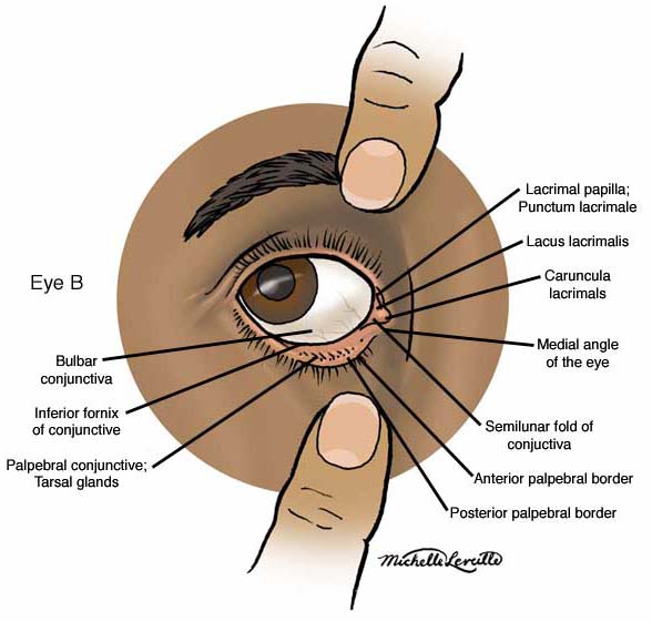
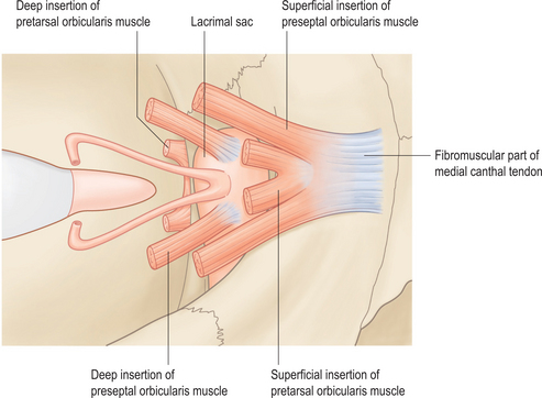
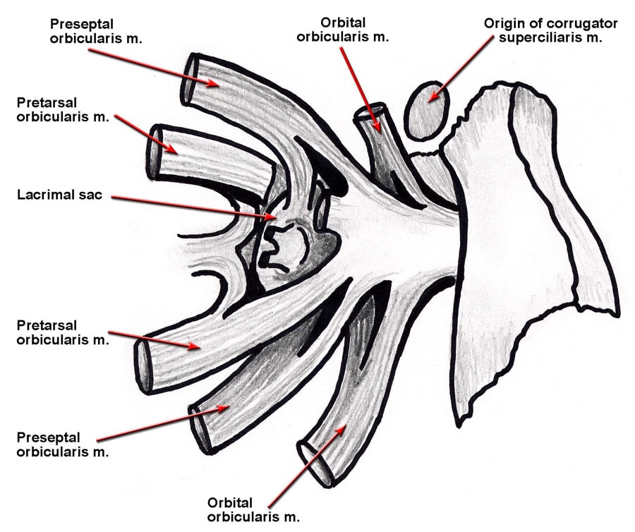
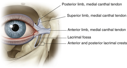
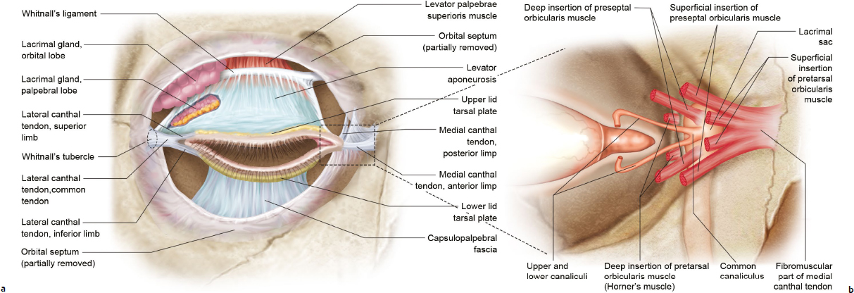
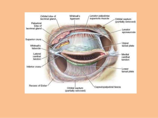
Medial canthus anatomy. The medial canthal tendon is formed by the merging of two tendinous arms originating from the anterior and posterior lacrimal crests. Any of several procedures for changing the configuration or position of the lateral canthus. The eyelids the nose is the inner canthus and the other is the outer canthus.
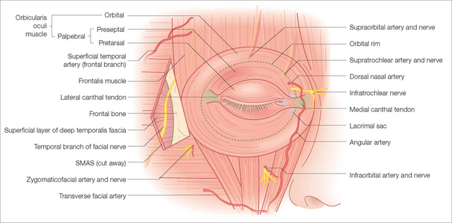
The upper and lower eyelids along with the upper and lower puncta oppose the globe. The medial palpebral ligament medial canthal tendon mct is a fibrous band stabilizing the medial tarsi and is intricately related with the orbicularis oculi muscle and the lacrimal system. Pinching the skin on the lower lip tests the mandibular branch.
Areas to be considered for full thickness grafts include the nasal ala the medial canthus of the eye the upper eyelid fingers and the ear. Touching the medial canthus of the eye evaluates the ophthalmic branch. More specifically the inner and outer canthi are respectively the medial and lateral endsangles of the palpebral fissure.
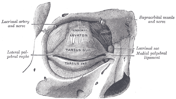
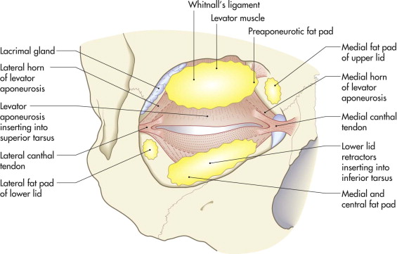
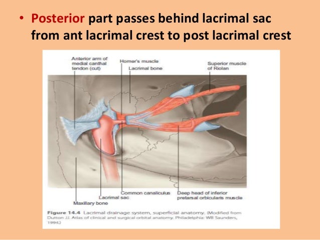
2 a muscular layer containing principally the orbicularis oculi muscle responsible for. Both of these have a unique angle at which the upper and lower eyelids meet. The superficial head of the pretarsal orbicularis muscle lies anterior to the canaliculi and forms the anterior limb of the mct.
Canthi palpebral commissures is either corner of the eye where the upper and lower eyelids meet. Laterally it is attached to the tarsus of the upper and lower eyelids. The structure of the palpebral fissure is maintained by the tarsal plates suspended by the medial and lateral canthal tendons fig.
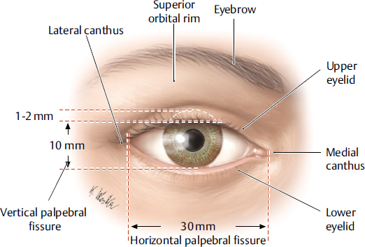
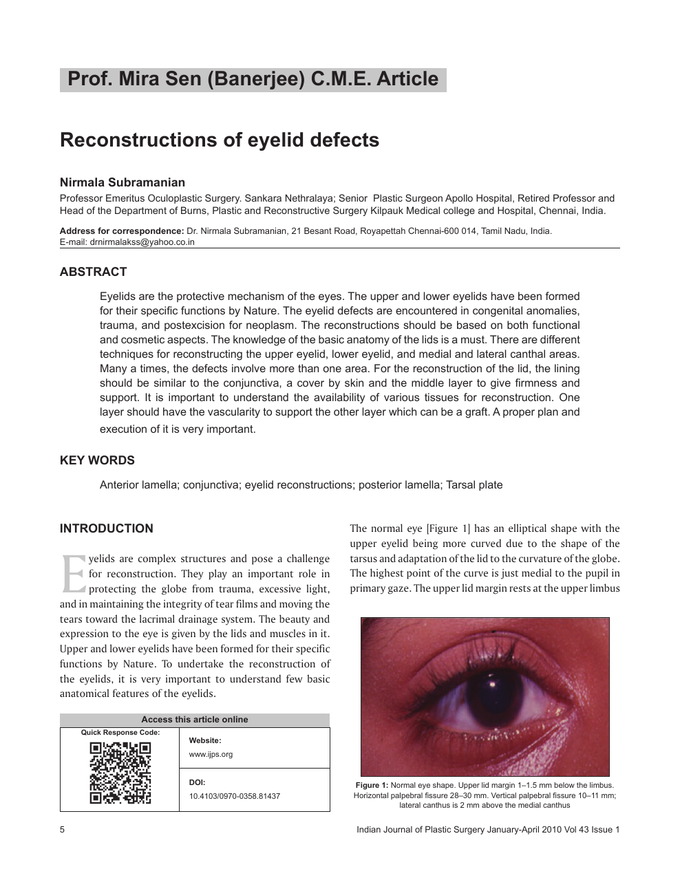
Used to correct deformities caused by trauma disease or prior surgery. The bicanthal plane is the transversal plane linking both canthi and defines the upper boundary of the midface. The upper lid naturally rests 1 to 2 mm below the superior limbus and peaks 1 mm medial to the center of the pupil.
The medial palpebral ligament medial canthal tendon is about 4 mm in length and 2 mm in breadth. Its anterior attachment is to the frontal process of the maxilla in front of the lacrimal groove and its posterior attachment is the lacrimal bone. A 75 year old male retired personnel presented with complaints of swelling at medial canthus of the left eye of one year duration associated with pain ocular discharge redness and watering.
When examined along a horizontal plane the medial canthal angle is located around 2 mm lower than the lateral canthal angle in caucasians. May also provide additional support to the lower eyelid by moving or tightening connections from the tarsal plate to the orbital rim. The lower lid rests at the inferior limbus and peaks 1 mm lateral to the center of the pupil.
The lid may be divided into four layers. 1 the skin containing glands that open onto the surface of the lid margin and the eyelashes.
 Lower Eyelid Surgery Vero Beach Lower Eyelid Surgery Melbourne
 Medial Palpebral Ligament Wikipedia
 Eyelid Disorders Diagnosis And Management American Family
 Blepharoplasty Plastic Surgery
 Repair Of The Lax Medial Canthal Tendon British Journal Of
 Eyelid Anatomy Plastic Surgery Key
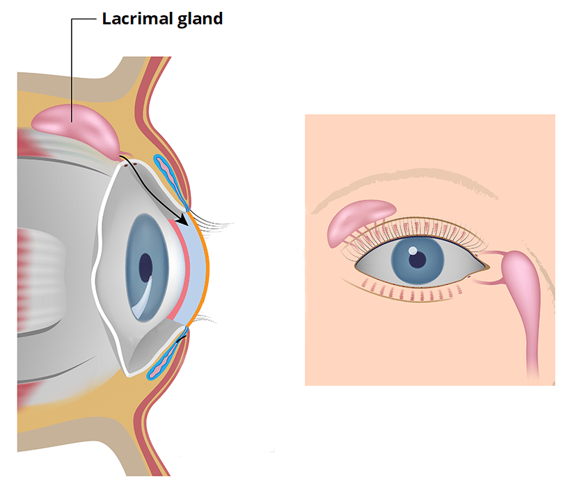 Lacrimal Glands And Apparatus Vasculature Innervation
 Anatomy Of The Eyelids
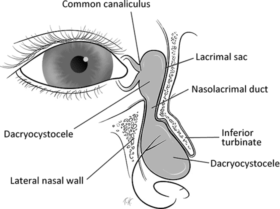 Congenital Dacryocystocele With Spontaneous Resolution
Anatomy Atlases Anatomy Of First Aid A Case Study Approach
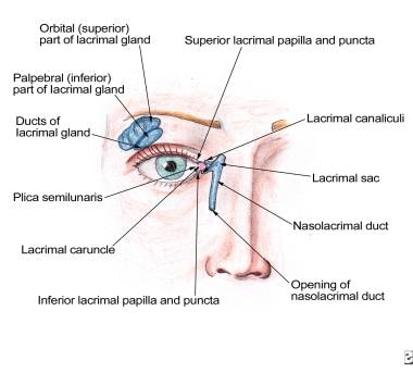 Nasolacrimal System Anatomy Embryology Puncta Canaliculi
 Observations In Ophthalmology Canine Eyelid Disease
 Eyelid And Midcheek Anatomy Plastic Surgery Key
 Periocular Reconstruction Clinical Gate
 Periocular Reconstruction Clinical Gate
 Eyelid And Lacrimal Injuries Pocket Dentistry
The Anatomy Of The Medial Canthal Ligament By T J Robinson
 Eyelid Anatomy Reconstruction And Blepharoplasty Plastic
 Ao Surgery Reference
 Anatomy Of The Eyelids
 Eyelid Anatomy Ento Key
 Lower Eyelid An Overview Sciencedirect Topics
 Eyelid Anatomy Plastic Surgery Key
 Reconstructive Options For The Medial Canthus And Eyelids
 Lower Eyelid And Eyelash Malpositions Ento Key
 Anatomy Of The Eyelids
 Closure Of Medial Canthus In Cats Procedure Efficacy
 Eyelid Anatomy For Cs Students Ppt Anatomy Of The Eyelids
 Caruncular Fixation In Medial Canthal Tendon Repair The Min
 Reconstructions Of Eyelid Defects Topic Of Research Paper
Belum ada Komentar untuk "Medial Canthus Anatomy"
Posting Komentar