Anatomy Of Esophagus And Trachea
Similarities between trachea and esophagus both trachea and esophagus are two tubular structures in the neck region of humans. The esophagus and the trachea are located at roughly the same place.
 Figure 2 From Surgical Anatomy Of The Trachea Semantic
The trachea is a median structure but near its lower end deviates slightly to the right resulting in the left main bronchus crossing anterior to the esophagus.
Anatomy of esophagus and trachea. The trachea descends anterior to the esophagus enters the superior mediastinum and divides into right and left main bronchi. A cricothyrotomy makes use of the easiest route of access through the cricothyroid ligament cricothyroid membrane between the cricoid cartilage below and thyroid cartilage above. Diagram esophagus and trachea see more about diagram esophagus and trachea diagram esophagus and trachea diagram showing esophagus and trachea diagram trachea and esophagus anatomy.
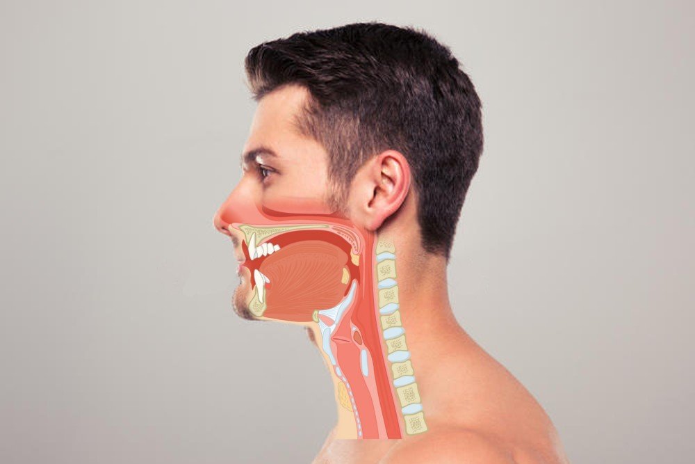
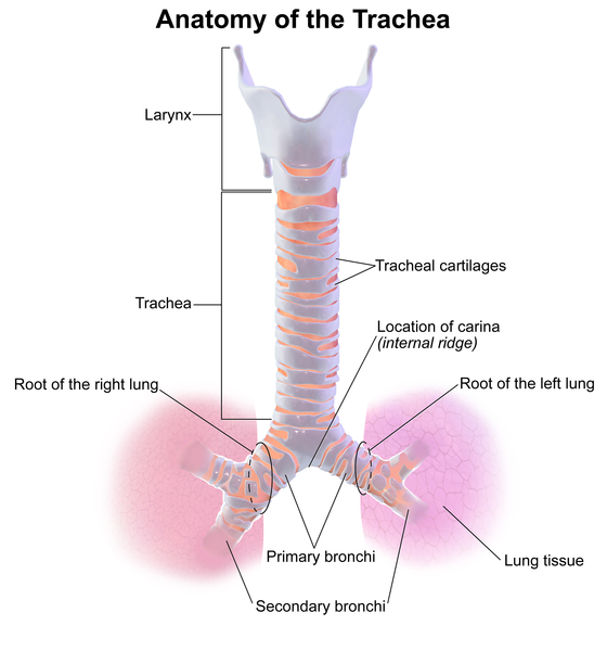
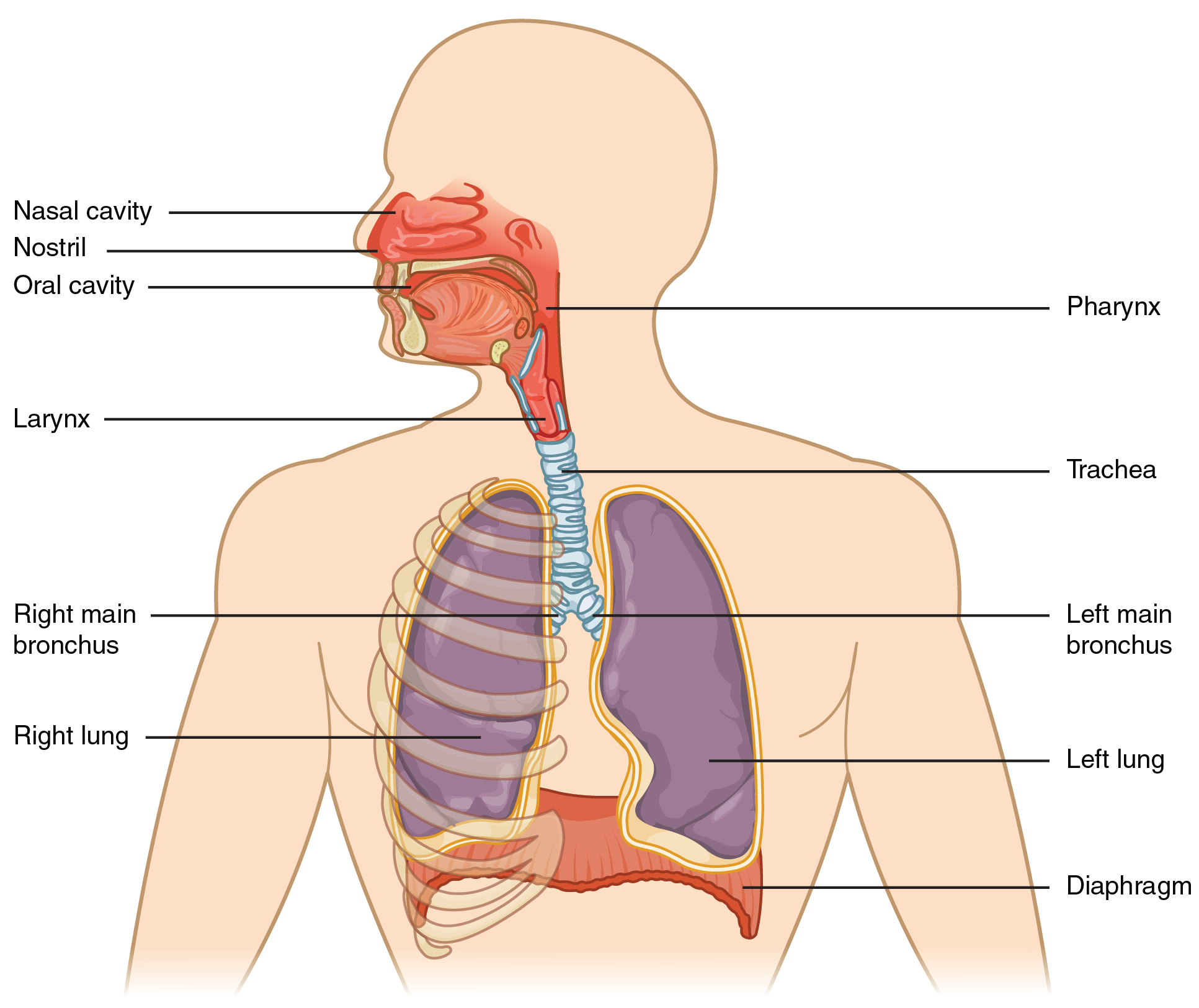
It is the link between your mouth and the stomach. The trachea lies behind the sternum breastbone and in front of the esophagus in the area of the chest between the lungs known as the mediastinum. The beginning of the trachea lies beneath the thyroid gland with the inferior end connecting with the carinathe area in which the main bronchus separates into two bronchi one of which enters each lung.
It divides into the left and right bronchus connected to the left and right lung respectively. The esophagus is a muscular tube connecting the throat pharynx with the stomach. Both trachea and esophagus are muscular tubes which are lined by a mucous membrane.
Muscular movements of the esophagus result in the passage of food from the mouth to the stomach cavity. The esophagus runs behind the windpipe trachea and heart and in front of the spine. Both trachea and esophagus perform transport functions.
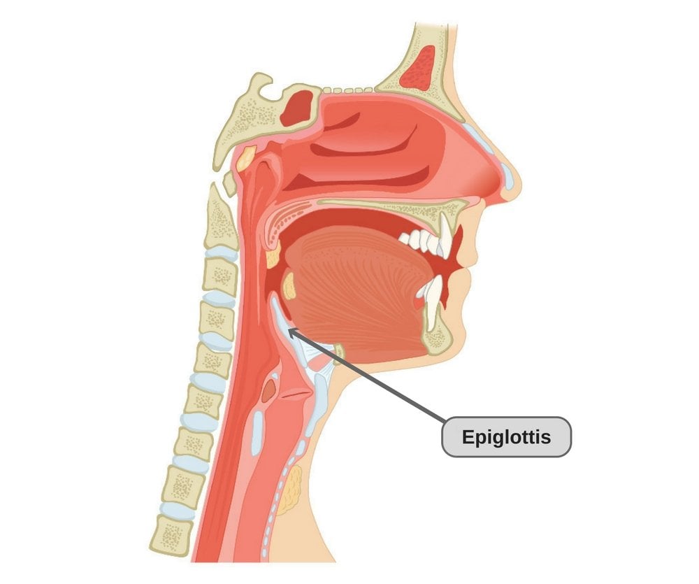
The esophagus is smaller and more flexible in structure naturally look at the amount of food it needs to transport. Beginning at the necks base just below the voice box the trachea is located in the thoracic or chest cavity in front of the esophagus running along the midline of the human body down to the back of the sternum breastbone. The larynx figure 2 and 4 and the trachea are anterior to the digestive tract esophagus in the neck and can be accessed directly when upper parts of the airway system are blocked.
Just before entering the stomach the esophagus passes through the diaphragm. The esophagus is about 8 inches long and is lined by moist pink tissue called mucosa.
 Heart Anatomy Model 7 Part Model Esophagus Trachea Svc Aorta Front Heart Wall Upper Half Of Heart
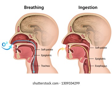
 What Are Esophagus And Trachea Why Are They Located Close
 Human Respiratory System Vector Illustration Cartoon Medical
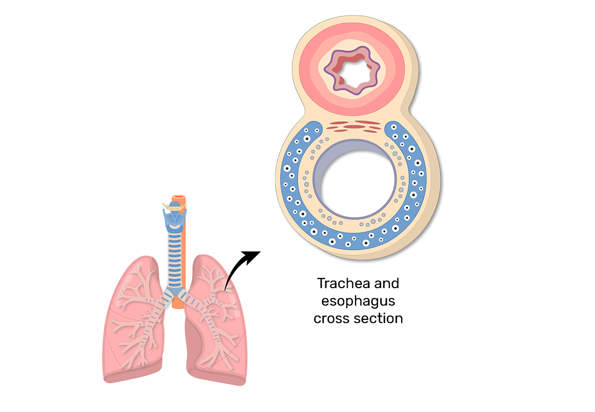
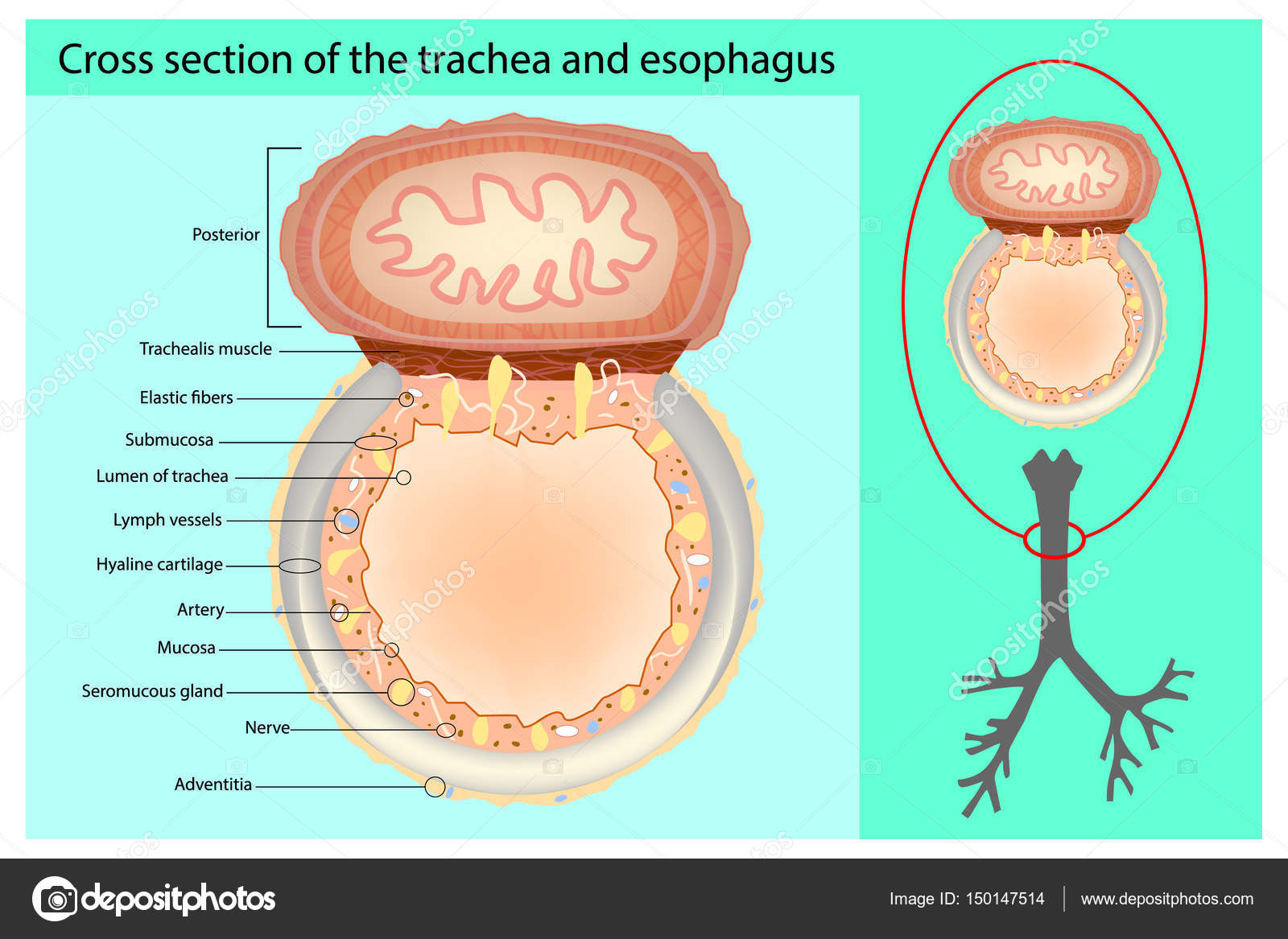 Picture Esophagus And Trachea Cross Section Of The
 Bronchus Wikipedia
 Throat Wikipedia
 General Surgery Thyroid Cancer
 What Are Esophagus And Trachea Why Are They Located Close
 Pin On Gross Anatomy
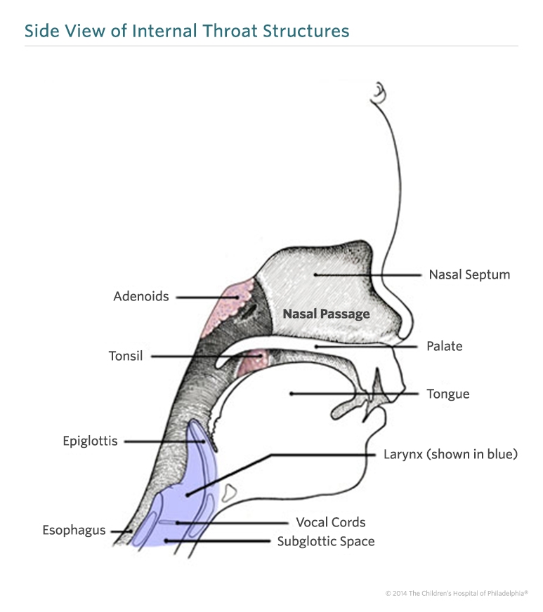 Throat Anatomy And Physiology Children S Hospital Of
 Esophagus Horse Health Simplified
Trachea Images Stock Photos Vectors Shutterstock

 Trachea Or Windpipe Location Anatomy And Physiology
 Ea Tef Genetics Home Reference Nih
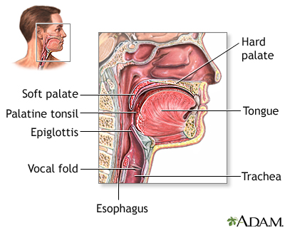 Throat Anatomy Medlineplus Medical Encyclopedia Image
 Trachea Images Stock Photos Vectors Shutterstock
 Diagram Of The Esophagus Diagram Of The Esophagus
 Trachea Windpipe Definition Anatomy Function Diagram
 Throat Pharyn Upper Esophageal Sphincter Ues Esophagus
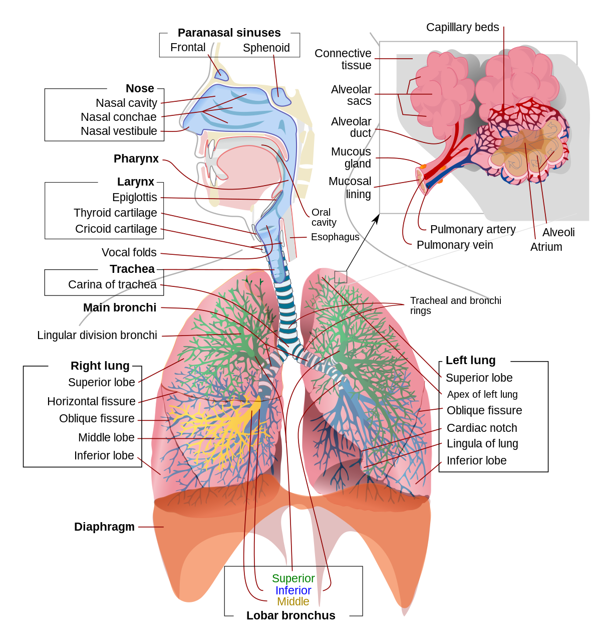
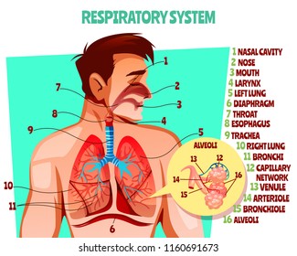
 22 1 Organs And Structures Of The Respiratory System
 Anatomy Of Esophagus Dr Neeraj Kumar Banoria

Is The Esophagus Posterior To The Trachea Quora
 What Are Esophagus And Trachea Why Are They Located Close
Belum ada Komentar untuk "Anatomy Of Esophagus And Trachea"
Posting Komentar